Names of Allah

Sunday, February 20, 2011

Tissue Culture and Horticulture

Although the idea of plant tissue and cell culture was conceived as early as 1902 by the German botanist Harberlandt, it was only during the last three decades or so that its application have increased considerably both in the basic research especially in the agriculture and horticulture. Now it has emerged as a multi-billion dollars industry for growing ornamentals and other botanicals.

Tissue Culture and Horticulture Starting from Himalaya foot hills to Karachi, Pakistan has a variety of ecological zones and a wide range of vegetation including flowers, foliage plants and crops which offers tremendous scope for exploiting tissueculture techniques for mass multiplication of economically important flora. This can be particularly useful in the production of economically important flora. This can be particularly useful in the production of elite and rare orchids and other plants, which have, export value. However, for effective utilisation of this technique it is necessary that scientists and private sector should interact and work on the practical application of the techniques for commercial purposes.

This has been successfully done in Malaysia, Srilanka and Singapore. Singapore alone is earning 2 million U.S. dollars by exporting of orchids mostly grown through this technique. Unfortunately we have not been able to exploit the tissue culture technique to a considerable extent. Time has now come to get into action to utilize the technology available for the benefit of the nation. There is yet no major commercial utilization of tissue culture in Pakistan. University of Agriculture can play a valuable role in the development of a program for the application of tissueculture in agriculture, horticulture, forestry, medicinal plants and ornamentals.

Tissue Culture and Horticulture Tissue culture technology is an established method of micropropagation of plants including cash crops, flowering shrubs and trees. Today nearly 50 companies in India are engaged in this business. This technology is particularly very useful for afforestation, as many trees take long time to grow and many of the conventional methods of propagation such as through seeds, cuttings and grafting are often slow and difficult.

Tissue culture is also useful for initial building-up of propagating stock of elite clones/individuals. It has found its best commercial application in propagation of clonal plants at very rapid rate compared to conventional methods. In certain cases it has been increased to million fold. Thus, it may immensely help in introducing newly created cultivars in trade. Presently, some 300 plant species have been demonstrated to be clonally propagatable through tissue culture (Murashige, 1978,1980; Vasil and Vasil, 1980; Flick et al., 1983). of the various plant species generally the ornamentals have been most successfully multiplied through tissue culture technique on commercial. The most glaring example is of orchid, the trade of which has been revolutionized by tissue culture (Hartmann and Kester, 1975; Morel, 1975; Rao, 1977).

History of Tissue Culture in Pakistan:

Hardly ten years ago the term plant tissue culture was totally new to most ofTissue Culture and Horticulture universities and research organizations in Pakistan. There were, however, sporadic reports from Department of Genetics, University of Karachi, C.C.R.I., Multan and Department of Botany University of Peshawar. From 1980 on ward there was a quantum leap in the number of tissue culture lab. in various parts of Pakistan although main activity in this field is conducted in public sector laboratories, we have now reached to a stage when the private sector is seemingly willing to invest in this new technology. Undoubtedly It has been emerged as a rapidly developing science because of its proven success on agriculture: in propagation, elimination of diseases, establishment of new varieties in a reduced span of time, shortened breeding cycle and conservation and propagation of endangered and threatened plant species.

Ability to establish and maintain plant organs (embryos, shoots, roots, flowers) and plant tissues (cells, callus, and protoplasts) in aseptic culture and to regenerate new plants from them is a result of basic and applied research in scientific laboratories (botany, plant pathology and genetics) since before the turn of the century. These separate procedures have been collectively called tissue and organ culture, in vitro culture, micropropagation, and most recently biotechnology. The commercial application of tissue culture was first discovered in the sixties in the west and since then it has become a multi-billion dollar industry for growing ornamental.

In coconut the biggest problem is the coconut root (wilt) disease and mycoplasm like organisms are involved. Tissue culture can come in handy to produce disease free planting material, which we require for large scale replanting.

Majority of commercial potato cultivar in uses e.g. Ultimus, Desiree, Malta, Cardinal and Diamont, are being imported every year in the form of virus free seed. The same seed could be prepared in this country by tissue culture techniques. (One example is the discovery of anther culture for haploid production which has become a potent tool for the plant breeder as well as for cell biologist. DR. Iqrar Ahmad Khan, Associate Professor, Department of Horticulture, University of Agriculture, Faisalabad). Plant tissue culture refers to in vitro and aseptic cultivation of plant part (cell or tissue) on a nutrient medium in an artificial environment.

Tissue Culture Methods

I. TYPES OF CELLS GROWN IN CULTURE
Tissue culture is often a generic term that refers to both organ culture and cell culture and the terms are often used interchangeably. Cell cultures are derived from either primary tissue explants or cell suspensions. Primary cell cultures typically will have a finite life span in culture whereas continuous cell lines are, by definition, abnormal and are often transformed cell lines.
II. WORK AREA AND EQUIPMENT
A. Laminar flow hoods. There are two types of laminar flow hoods, vertical and horizontal. The vertical hood, also known as a biology safety cabinet, is best for working with hazardous organisms since the aerosols that are generated in the hood are filtered out before they are released into the surrounding environment. Horizontal hoods are designed such that the air flows directly at the operator hence they are not useful for working with hazardous organisms but are the best protection for your cultures. Both types of hoods have continuous displacement of air that passes through a HEPA (high efficiency particle) filter that removes particulates from the air. In a vertical hood, the filtered air blows down from the top of the cabinet; in a horizontal hood, the filtered air blows out at the operator in a horizontal fashion. NOTE: these are not fume hoods and should not be used for volatile or explosive chemicals. They should also never be used for bacterial or fungal work. The hoods are equipped with a short-wave UV light that can be turned on for a few minutes to sterilize the surfaces of the hood, but be aware that only exposed surfaces will be accessible to the UV light. Do not put your hands or face near the hood when the UV light is on as the short wave light can cause skin and eye damage. The hoods should be turned on about 10-20 minutes before being used. Wipe down all surfaces with ethanol before and after each use. Keep the hood as free of clutter as possible because this will interfere with the laminar flow air pattern.
B. CO2 Incubators. The cells are grown in an atmosphere of 5-10% CO2 because the medium used is buffered with sodium bicarbonate/carbonic acid and the pH must be strictly maintained. Culture flasks should have loosened caps to allow for sufficient gas exchange. Cells should be left out of the incubator for as little time as possible and the incubator doors should not be opened for very long. The humidity must also be maintained for those cells growing in tissue culture dishes so a pan of water is kept filled at all times.
C. Microscopes. Inverted phase contrast microscopes are used for visualizing the cells. Microscopes should be kept covered and the lights turned down when not in use. Before using the microscope or whenever an objective is changed, check that the phase rings are aligned.
D. Preservation. Cells are stored in liquid nitrogen (see Section III- Preservation and storage).
E. Vessels. Anchorage dependent cells require a nontoxic, biologically inert, and optically transparent surface that will allow cells to attach and allow movement for growth. The most convenient vessels are specially-treated polystyrene plastic that are supplied sterile and are disposable. These include petri dishes, multi-well plates, microtiter plates, roller bottles, and screwcap flasks - T-25, T-75, T-150 (cm2 of surface area). Suspension cells are either shaken, stirred, or grown in vessels identical to those used for anchorage-dependent cells.
III. PRESERVATION AND STORAGE. Liquid N2 is used to preserve tissue culture cells, either in the liquid phase (-196°C) or in the vapor phase (-156°C). Freezing can be lethal to cells due to the effects of damage by ice crystals, alterations in the concentration of electrolytes, dehydration, and changes in pH. To minimize the effects of freezing, several precautions are taken. First, a cryoprotective agent which lowers the freezing point, such as glycerol or DMSO, is added. A typical freezing medium is 90% serum, 10% DMSO. In addition, it is best to use healthy cells that are growing in log phase and to replace the medium 24 hours before freezing. Also, the cells are slowly cooled from room temperature to -80°C to allow the water to move out of the cells before it freezes. The optimal rate of cooling is 1°-3°C per minute. Some labs have fancy freezing chambers to regulate the freezing at the optimal rate by periodically pulsing in liquid nitrogen. We use a low tech device called a Mr. Frosty (C#1562 -Nalgene, available from Sigma). The Mr. Frosty is filled with 200 ml of isopropanol at room temperature and the freezing vials containing the cells are placed in the container and the container is placed in the -80°C freezer. The effect of the isopropanol is to allow the tubes to come to the temperature of the freezer slowly, at about 1°C per minute. Once the container has reached -80°C (about 4 hours or, more conveniently, overnight) the vials are removed from the Mr. Frosty and immediately placed in the liquid nitrogen storage tank. Cells are stored at liquid nitrogen temperatures because the growth of ice crystals is retarded below -130°C. To maximize recovery of the cells when thawing, the cells are warmed very quickly by placing the tube directly from the liquid nitrogen container into a 37°C water bath with moderate shaking. As soon as the last ice crystal is melted, the cells are immediately diluted into prewarmed medium.
IV. MAINTENANCE
Cultures should be examined daily, observing the morphology, the color of the medium and the density of the cells. A tissue culture log should be maintained that is separate from your regular laboratory notebook. The log should contain: the name of the cell line, the medium components and any alterations to the standard medium, the dates on which the cells were split and/or fed, a calculation of the doubling time of the culture (this should be done at least once during the semester), and any observations relative to the morphology, etc.
A. Growth pattern. Cells will initially go through a quiescent or lag phase that depends on the cell type, the seeding density, the media components, and previous handling. The cells will then go into exponential growth where they have the highest metabolic activity. The cells will then enter into stationary phase where the number of cells is constant, this is characteristic of a confluent population (where all growth surfaces are covered).
B. Harvesting. Cells are harvested when the cells have reached a population density which suppresses growth. Ideally, cells are harvested when they are in a semi-confluent state and are still in log phase. Cells that are not passaged and are allowed to grow to a confluent state can sometime lag for a long period of time and some may never recover. It is also essential to keep your cells as happy as possible to maximize the efficiency of transformation. Most cells are passaged (or at least fed) three times a week.
1. Suspension culture. Suspension cultures are fed by dilution into fresh medium.
2. Adherent cultures. Adherent cultures that do not need to be divided can simply be fed by removing the old medium and replacing it with fresh medium.
When the cells become semi-confluent, several methods are used to remove the cells from the growing surface so that they can be diluted:
  • Mechanical - A rubber spatula can be used to physically remove the cells from the growth surface. This method is quick and easy but is also disruptive to the cells and may result in significant cell death. This method is best when harvesting many different samples of cells for preparing extracts, i.e., when viability is not important.
  • Proteolytic enzymes - Trypsin, collagenase, or pronase, usually in combination with EDTA, causes cells to detach from the growth surface. This method is fast and reliable but can damage the cell surface by digesting exposed cell surface proteins. The proteolysis reaction can be quickly terminated by the addition of complete medium containing serum
  • EDTA - EDTA alone can also be used to detach cells and seems to be gentler on the cells than trypsin. The standard procedure for detaching adherent cells is as follows:
    1. Visually inspect daily 2. Release cells from monolayer surface
  a. wash once with a buffer solution
b. treat with dissociating agent
c. observe cells under the microscope. Incubate until cells become rounded and loosen when flask is gently tapped with the side of the hand.
d. Transfer cells to a culture tube and dilute with medium containing serum.
e. Spin down cells, remove supernatant and replace with fresh medium.
f. Count the cells in a hemacytometer, and dilute as appropriate into fresh medium.
C. Media and growth requirements
1. Physiological parameters
A. temperature - 37C for cells from homeother
B. pH - 7.2-7.5 and osmolality of medium must be maintained
C. humidity is required
D. gas phase - bicarbonate conc. and CO2 tension in equilibrium
E. visible light - can have an adverse effect on cells; light induced production of toxic compounds can occur in some media; cells should be cultured in the dark and exposed to room light as little as possible;
2. Medium requirements: (often empirical)
A. Bulk ions - Na, K, Ca, Mg, Cl, P, Bicarb or CO2
B. Trace elements - iron, zinc, selenium
C. sugars - glucose is the most common
D. amino acids - 13 essential
E. vitamins - B, etc.
F. choline, inositol
G. serum - contains a large number of growth promoting activities such as buffering toxic nutrients by binding them, neutralizes trypsin and other proteases, has undefined effects on the interaction between cells and substrate, and contains peptide hormones or hormone-like growth factors that promote healthy growth.
H. antibiotics - although not required for cell growth, antibiotics are often used to control the growth of bacterial and fungal contaminants.

3. Feeding - 2-3 times/week.
4. Measurement of growth and viability. The viability of cells can be observed visually using an inverted phase contrast microscope. Live cells are phase bright; suspension cells are typically rounded and somewhat symmetrical; adherent cells will form projections when they attach to the growth surface. Viability can also be assessed using the vital dye, trypan blue, which is excluded by live cells but accumulates in dead cells. Cell numbers are determined using a hemocytometer.
V. SAFETY CONSIDERATIONS
bullet Assume all cultures are hazardous since they may harbor latent viruses or other organisms that are uncharacterized. The following safety precautions should also be observed:
bullet pipetting: use pipette aids to prevent ingestion and keep aerosols down to a minimum
bullet no eating, drinking, or smoking
bullet wash hands after handling cultures and before leaving the lab
bullet decontaminate work surfaces with disinfectant (before and after)
bullet autoclave all waste
bullet use biological safety cabinet (laminar flow hood) when working with hazardous organisms. The cabinet protects worker by preventing airborne cells and viruses released during experimental activity from escaping the cabinet; there is an air barrier at the front opening and exhaust air is filtered with a HEPA filter make sure cabinet is not overloaded and leave exhaust grills in the front and the back clear (helps to maintain a uniform airflow)
bullet use aseptic technique
bullet dispose of all liquid waste after each experiment and treat with bleach

REFERENCES:
R. Ian Freshney, Culture of Animal cells: A manual of basic techniques, Wiley-Liss, 1987.
VI. TISSUE CULTURE PROCEDURES
Each student should maintain his own cells throughout the course of the experiment. These cells should be monitored daily for morphology and growth characteristics, fed every 2 to 3 days, and subcultured when necessary. A minimum of two 25 cm2 flasks should be carried for each cell line; these cells should be expanded as necessary for the transfection experiments. Each time the cells are subcultured, a viable cell count should be done, the subculture dilutions should be noted, and, after several passages, a doubling time determined. As soon as you have enough cells, several vials should be frozen away and stored in liquid N2. One vial from each freeze down should be thawed 1-2 weeks after freezing to check for viability. These frozen stocks will prove to be vital if any of your cultures become contaminated.
Procedures:1. Media preparation. Each student will be responsible for maintaining his own stock of cell culture media; the particular type of media, the sera type and concentration, and other supplements will depend on the cell line. Do not share media with you partner (or anyone else) because if a culture or a bottle of media gets contaminated, you have no back-up. Most of the media components will be purchased prepared and sterile. In general, all you need to do is sterily combine several sterile solutions. To test for sterility after adding all components, pipet several mls from each media bottle into a small sterile petri dish or culture tube and incubate at 37EC for several days. Use only media that has been sterility tested. For this reason, you must anticipate your culture needs in advance so you can prepare the reagents necessary. But, please try not to waste media. Anticipate your needs but don't make more than you need. Tissue culture reagents are very expensive; for example, bovine fetal calf serum cost ~ $200/500 ml. Some cell culture additives will be provided in a powdered form. These should be reconstituted to the appropriate concentration with double-distilled water (or medium, as appropriate) and filtered (in a sterile hood) through a 0-22 μm filter.
All media preparation and other cell culture work must be performed in a laminar flow hood. Before beginning your work, turn on blower for several minutes, wipe down all surfaces with 70% ethanol, and ethanol wash your clean hands. Use only sterile pipets, disposable test tubes and autoclaved pipet tips for cell culture. All culture vessels, test tubes, pipet tip boxes, stocks of sterile eppendorfs, etc. should be opened only in the laminar flow hood. If something is opened elsewhere in the lab by accident, you can probably assume its contaminated. If something does become contaminated, immediately discard the contaminated materials into the biohazard container and notify the instructor.
2. Growth and morphology. Visually inspect cells frequently. Cell culture is sometimes more an art than a science. Get to know what makes your cells happy. Frequent feeding is important for maintaining the pH balance of the medium and for eliminating waste products. Cells do not typically like to be too confluent so they should be subcultured when they are in a semi-confluent state. In general, mammalian cells should be handled gently. They should not be vortexed, vigorously pipetted or centrifuged at greater than 1500 g.
3. Cell feeding. Use prewarmed media and have cells out of the incubator for as little time as possible. Use 10-15 ml for T-25's, 25-35 ml for T-75's and 50-60 ml for T-150's. a. Suspension cultures. Feeding and subculturing suspension cultures are done simultaneously. About every 2-3 days, dilute the cells into fresh media. The dilution you use will depend on the density of the cells and how quickly they divide, which only you can determine. Typically 1:4 to 1:20 dilutions are appropriate for most cell lines. b. Adherent cells. About every 2-3 days, pour off old media from culture flasks and replace with fresh media. Subculture cells as described below before confluency is reached.
4. Subculturing adherent cells. When adherent cells become semi-confluent, subculture using 2 mM EDTA or trypsin/EDTA.
Trypsin-EDTA :
bullet a. Remove medium from culture dish and wash cells in a balanced salt solution without Ca++ or Mg++. Remove the wash solution.
bullet b. Add enough trypsin-EDTA solution to cover the bottom of the culture vessel and then pour off the excess.
bullet c. Place culture in the 37°C incubator for 2 minutes.
bullet d. Monitor cells under microscope. Cells are beginning to detach when they appear rounded.
bullet e. As soon as cells are in suspension, immediately add culture medium containing serum. Wash cells once with serum containing medium and dilute as appropriate (generally 4-20 fold).
EDTA alone:
bullet a. Prepare a 2 mM EDTA solution in a balanced salt solution (i.e., PBS without Ca++ or Mg++).
bullet b. Remove medium from culture vessel by aspiration and wash the monolayer to remove all traces of serum. Remove salt solution by aspiration.
bullet c. Dispense enough EDTA solution into culture vessels to completely cover the monolayer of cells.
bullet d. The coated cells are allowed to incubate until cells detach from the surface. Progress can be checked by examination with an inverted microscope. Cells can be gently nudged by banging the side of the flask against the palm of the hand.
bullet e. Dilute cells with fresh medium and transfer to a sterile centrifuge tube.
bullet f. Spin cells down, remove supernatant, and resuspend in culture medium (or freezing medium if cells are to be frozen). Dilute as appropriate into culture flasks.
5. Thawing frozen cells.
bullet a. Remove cells from frozen storage and quickly thaw in a 37°C waterbath by gently agitating vial.
bullet b. As soon as the ice crystals melt, pipet gently into a culture flask containing prewarmed growth medium.
bullet c. Log out cells in the "Liquid Nitrogen Freezer Log" Book.
6. Freezing cells.
bullet a. Harvest cells as usual and wash once with complete medium.
bullet b. Resuspend cells in complete medium and determine cell count/viability.
bullet c. Centrifuge and resuspend in ice-cold freezing medium: 90% calf serum/10% DMSO, at 106 - 107 cells/ml. Keep cells on ice.
bullet d. Transfer 1 ml aliquots to freezer vials on ice.
bullet e. Place in a Mr. Frosty container that is at room temperature and that has sufficient isopropanol.
bullet f. Place the Mr. Frosty in the -70°C freezer overnight. Note: Cells should be exposed to freezing medium for as little time as possible prior to freezing
bullet g Next day, transfer to liquid nitrogen (DON'T FORGET) and log in the "Liquid Nitrogen Freezer Log" Book.
7. Viable cell counts. USING A HEMOCYTOMETER TO DETERMINE TOTAL CELL COUNTS AND VIABLE CELL NUMBERS (Reference: Sigma catalogue)Trypan blue is one of several stains recommended for use in dye exclusion procedures for viable cell counting. This method is based on the principle that live cells do not take up certain dyes, whereas dead cells do.
1. Prepare a cell suspension, either directly from a cell culture or from a concentrated or diluted suspension (depending on the cell density) and combine 20 μl of cells with 20 μl of trypan blue suspension (0.4%). Mix thoroughly and allow to stand for 5-15 minutes.
2. With the cover slip in place, transfer a small amount of trypan blue-cell suspension to both chambers of the hemocytometer by carefully touching the edge of the cover slip with the pipette tip and allowing each chamber to fill by capillary action. Do not overfill or underfill the chambers.3. Starting with 1 chamber of the hemocytometer, count all the cells in the 1 mm center square and four 1 mm corner square.  Keep a separate count of viable and non-viable cells.4. If there are too many or too few cells to count, repeat the procedure either concentrating or diluting the original suspension as appropriate.5. The circle indicates the approximate area covered at 100X microscope magnification (10X ocular and 10X objective). Include cells on top and left touching middle line. Do not count cells touching middle line at bottom and right. Count 4 corner squares and middle square in both chambers and calculate the average.6. Each large square of the hemocytometer, with cover-slip in place, represents a total volume of 0.1 mm3 or 10-4 cm3. Since 1 cm3 is equivalent to approximately 1 ml, the total number of cells per ml will be determined using the following calculations:Cells/ml = average cell count per square x dilution factor x 104;
Total cells = cells/ml x the original volume of fluid from which the cell sample was removed; % Cell viability = total viable cells (unstained)/total cells x 100.

Plant Tissue Culture

Plant tissue culture is a practice used to propagate plants under sterile conditions, often to produce clones of a plant. Different techniques in plant tissue culture may offer certain advantages over traditional methods of propagation, including:
  • The production of exact copies of plants that produce particularly good flowers, fruits, or have other desirable traits.
  • To quickly produce mature plants.
  • The production of multiples of plants in the absence of seeds or necessary pollinators to produce seeds.
  • The regeneration of whole plants from plant cells that have been genetically modified.
  • The production of plants in sterile containers that allows them to be moved with greatly reduced chances of transmitting diseases, pests, and pathogens.
  • The production of plants from seeds that otherwise have very low chances of germinating and growing, i.e.: orchids and nepenthes.
  • To clean particular plant of viral and other infections and to quickly multiply these plants as 'cleaned stock' for horticulture and agriculture.
Plant tissue culture relies on the fact that many plant cells have the ability to regenerate a whole plant (totipotency). Single cells, plant cells without cell walls (protoplasts), pieces of leaves, or (less commonly) roots can often be used to generate a new plant on culture media given the required nutrients and plant hormones.

Contents

  • 1 Techniques
  • 2 Choice of explant
  • 3 Applications
  • 4 Laboratories

Techniques

Modern plant tissue culture is performed under aseptic conditions under filtered air. Living plant materials from the environment are naturally contaminated on their surfaces (and sometimes interiors) with microorganisms, so surface sterilization of starting materials (explants) in chemical solutions (usually alcohol or bleach) is required. Mercuric chloride is seldom used as a plant sterilant today, as it is dangerous to use, and is difficult to dispose of. Explants are then usually placed on the surface of a solid culture medium, but are sometimes placed directly into a liquid medium, particularly when cell suspension cultures are desired. Solid and liquid media are generally composed of inorganic salts plus a few organic nutrients, vitamins and plant hormones. Solid media are prepared from liquid media with the addition of a gelling agent, usually purified agar.
In-vitro tissue culture potato explants
The composition of the medium, particularly the plant hormones and the nitrogen source (nitrate versus ammonium salts or amino acids) have profound effects on the morphology of the tissues that grow from the initial explant. For example, an excess of auxin will often result in a proliferation of roots, while an excess of cytokinin may yield shoots. A balance of both auxin and cytokinin will often produce an unorganised growth of cells, or callus, but the morphology of the outgrowth will depend on the plant species as well as the medium composition. As cultures grow, pieces are typically sliced off and transferred to new media (subcultured) to allow for growth or to alter the morphology of the culture. The skill and experience of the tissue culturist are important in judging which pieces to culture and which to discard.
As shoots emerge from a culture, they may be sliced off and rooted with auxin to produce plantlets which, when mature, can be transferred to potting soil for further growth in the greenhouse as normal plants.[1]

Choice of explant

The tissue obtained from the plant to culture is called an explant. Based on work with certain model systems, particularly tobacco, it has often been claimed that a totipotent explant can be grown from any part of the plant. However, this concept has been vitiated in practice. In many species explants of various organs vary in their rates of growth and regeneration, while some do not grow at all. The choice of explant material also determines if the plantlets developed via tissue culture are haploid or diploid. Also the risk of microbial contamination is increased with inappropriate explants. Thus it is very important that an appropriate choice of explant be made prior to tissue culture.
The specific differences in the regeneration potential of different organs and explants have various explanations. The significant factors include differences in the stage of the cells in the cell cycle, the availability of or ability to transport endogenous growth regulators, and the metabolic capabilities of the cells. The most commonly used tissue explants are the meristematic ends of the plants like the stem tip, auxiliary bud tip and root tip. These tissues have high rates of cell division and either concentrate or produce required growth regulating substances including auxins and cytokinins.
Some explants, like the root tip, are hard to isolate and are contaminated with soil microflora that become problematic during the tissue culture process. Certain soil microflora can form tight associations with the root systems, or even grow within the root. Soil particles bound to roots are difficult to remove without injury to the roots that then allows microbial attack. These associated microflora will generally overgrow the tissue culture medium before there is significant growth of plant tissue.
Aerial (above soil) explants are also rich in undesirable microflora. However, they are more easily removed from the explant by gentle rinsing, and the remainder usually can be killed by surface sterilization. Most of the surface microflora do not form tight associations with the plant tissue. Such associations can usually be found by visual inspection as a mosaic, de-colorization or localized necrosis on the surface of the explant.
An alternative for obtaining uncontaminated explants is to take explants from seedlings which are aseptically grown from surface-sterilized seeds. The hard surface of the seed is less permeable to penetration of harsh surface sterilizing agents, such as hypochlorite, so the acceptable conditions of sterilization used for seeds can be much more stringent than for vegetative tissues.
Tissue cultured plants are clones, if the original mother plant used to produce the first explants is susceptible to a pathogen or environmental condition, the entire crop would be susceptible to the same problem, conversely any positive traits would remain within the line also.

Applications

Plant tissue culture is used widely in plant science; it also has a number of commercial applications. Applications include:
  • Micropropagation is widely used in forestry and in floriculture. Micropropagation can also be used to conserve rare or endangered plant species.
  • A plant breeder may use tissue culture to screen cells rather than plants for advantageous characters, e.g. herbicide resistance/tolerance.
  • Large-scale growth of plant cells in liquid culture in bioreactors for production of valuable compounds, like plant-derived secondary metabolites and recombinant proteins used as biopharmaceuticals [2].
  • To cross distantly related species by protoplast fusion and regeneration of the novel hybrid.
  • To cross-pollinate distantly related species and then tissue culture the resulting embryo which would otherwise normally die (Embryo Rescue).
  • For production of doubled monoploid (dihaploid) plants from haploid cultures to achieve homozygous lines more rapidly in breeding programmes, usually by treatment with colchicine which causes doubling of the chromosome number.
  • As a tissue for transformation, followed by either short-term testing of genetic constructs or regeneration of transgenic plants.
  • Certain techniques such as meristem tip culture can be used to produce clean plant material from virused stock, such as potatoes and many species of soft fruit.
  • micropropagation using meristem and shoot culture to produce large numbers of identical individuals.

Laboratories

Although some growers and nurseries have their own labs for propagating plants by the technique of tissue culture, a number of independent laboratories provide custom propagation services. The Plant Tissue Culture Information Exchange lists many commercial tissue culture labs. Since plant tissue culture is a very labour intensive process, this would be an important factor in determining which plants would be commercially viable to propagate in a laboratory.

Tissue culture

Flasks containing tissue culture growth medium which provides nourishment to growing cells.
Tissue culture is the growth of tissues and/or cells separate from the organism. This is typically facilitated via use of a liquid, semi-solid, or solid growth medium, such as broth or agar. Tissue culture commonly refers to the culture of animal cells and tissues, while the more specific term plant tissue culture is being named for the plants.

Contents

  • 1 Historical usage
  • 2 Modern usage

Historical usage

In 1885 Wilhelm Roux removed a section of the medullary plate of an embryonic chicken and maintained it in a warm saline solution for several days, establishing the basic principle of tissue culture.
In 1907 the zoologist Ross Granville Harrison demonstrated the growth of frog nerve cell processes in a medium of clotted lymph.
In 1913, E. Steinhardt, C. Israeli, and R. A. Lambert grew vaccinia virus in fragments of guinea pig corneal tissue.[1]

Modern usage

In modern usage, "tissue culture" generally refers to the growth of eukaryotic cells in vitro. It is often used interchangeably with cell culture to specifically describe the in vitro culturing of sperm donor cells.
However, "tissue culture" can also be used to refer to the culturing of tissue pieces, i.e. explant culture or whole organs, i.e. organ culture.
It is a tool for the study of animal cell biology in vitro model of cell growth to allow a highly selective environment which is easily manipulated (used to optimize cell signaling pathways).

One Amazing Dish

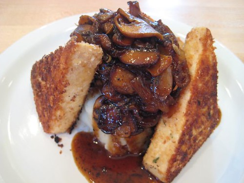
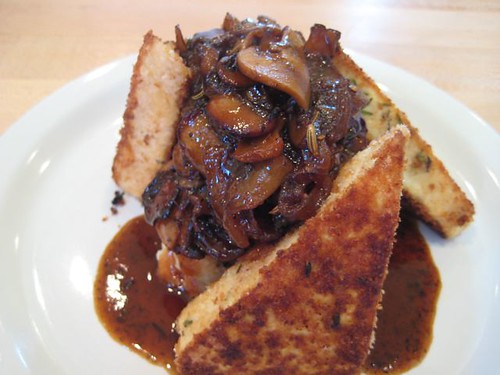
Let me start off by saying, this is not your 70's-style tofu. This is tofu prepared in an elegant and upscale way. This is fine dining tofu.

Imagine Marsala-marinated tofu, coated with herbed panko crumbs and pan-seared until it's got a gorgeous crispy golden crust. Close your eyes and imagine this ethereal tofu perched on a mound of rosemary garlic mashed potatoes, and topped with an earthy mushroom Marsala sauce. Now this is tofu.

I first tasted this tofu dish at The Farm Cafe in Portland Oregon. I was having dinner with my good buddy Joanna, and she highly recommended the tofu. After careful scanning of the menu, chock-full of local, fresh, organic produce, this was definitely the dish that caught my eye. Herbed Crusted Tofu With A Mushroom Marsala Sauce. OMG! It had my name written all over it! The dish was divine. So much so that I started holding it up as my standard of what a good tofu dish should be. No problem preparing this at home, right? Wrong, actually. This was a tougher nut to crack than I imagined.

So, fast forward to last month, when the owner of The Farm Cafe accepted my offer to come prepare her amazing herbed-crusted tofu on Everyday Dish. Now everyone can learn their secret to a fabulously prepared tofu dish. And, we all have a delicious new tofu recipe to serve at our next dinner party, or try out on our tofu-phobic husbands/children/friends.

The recipe and video are up now on Everyday Dish. Thank you Fearn and Russel and The Farm Cafe, for sharing your delicious recipe with us. Now, what are you waiting for? You've got to try this dish!

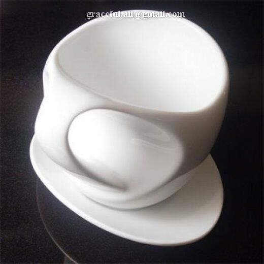
Amazing Designer Cups

Amazing Cups

Amazing Designer Cups

Amazing Designer Cups

Amazing Designer Cups

Amazing Designer Cups

Amazing Designer Cups

Amazing Designer Cups

Amazing Designer Cups

Amazing Designer Cups

Amazing Designer Cups

Amazing Designer Cups

Amazing Designer Cups

Amazing Designer Cups