
The brain is the center of the nervous system in all vertebrate and most invertebrate animals.[1] Some primitive animals such as jellyfish and starfish have a decentralized nervous system without a brain, while sponges lack any nervous system at all. In vertebrates the brain is located in the head, protected by the skull and close to the primary sensory apparatus of vision, hearing, balance, taste, and smell.
Brains can be extremely complex. The cerebral cortex of the human brain contains roughly 15–33 billion neurons, perhaps more, depending on gender and age,[2] linked with up to 10,000 synaptic connections each. Each cubic millimeter of cerebral cortex contains roughly one billion synapses.[3] These neurons communicate with one another by means of long protoplasmic fibers called axons, which carry trains of signal pulses called action potentials to distant parts of the brain or body and target them to specific recipient cells.
The brain controls the other organ systems of the body, either by activating muscles or by causing secretion of chemicals such as hormones and neurotransmitters. This centralized control allows rapid and coordinated responses to changes in the environment. Some basic types of responsiveness are possible without a brain: even single-celled organisms may be capable of extracting information from the environment and acting in response to it.[4] Sponges, which lack a central nervous system, are capable of coordinated body contractions and even locomotion.[5] In vertebrates, the spinal cord by itself contains neural circuitry capable of generating reflex responses as well as simple motor patterns such as swimming or walking.[6] However, sophisticated control of behavior on the basis of complex sensory input requires the information-integrating capabilities of a centralized brain.
Despite rapid scientific progress, much about how brains work remains a mystery. The operations of individual neurons and synapses are now understood in considerable detail, but the way they cooperate in ensembles of thousands or millions has been very difficult to decipher. Methods of observation such as EEG recording and functional brain imaging tell us that brain operations are highly organized, while single unit recording can resolve the activity of single neurons, but how individual cells give rise to complex operations is unknown.[7]


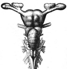
 Nervous system of a bilaterian animal, in the form of a nerve cord with segmental enlargements, and a "brain" at the frontWith the exception of a few primitive forms such as sponges and jellyfish, all living animals are bilateria, meaning animals with a bilaterally symmetric body shape (that is, left and right sides that are approximate mirror images of each other).[9]
All bilateria are thought to have descended from a common ancestor that appeared early in the Cambrian period, 550–600 million years ago.[9] This ancestor had the shape of a simple tube worm with a segmented body, and at an abstract level, that worm-shape continues to be reflected in the body and nervous system plans of all modern bilateria, including humans.[10] The fundamental bilateral body form is a tube with a hollow gut cavity running from mouth to anus, and a nerve cord with an enlargement (a "ganglion") for each body segment, with an especially large ganglion at the front, called the "brain".
There are a few invertebrates whose brains have been studied intensively. The large sea slug Aplysia was chosen by Nobel Prize-winning neurophysiologist Eric Kandel, because of the simplicity and accessibility of its nervous system, as a model for studying the cellular basis of learning and memory, and subjected to hundreds of experiments.[12] The most thoroughly studied invertebrate brains, however, belong to the fruit fly Drosophila and the tiny roundworm Caenorhabditis elegans (C. elegans).
Because of the large array of techniques available for studying their genetics, fruit flies have been a natural subject for studying the role of genes in brain development.[13] Remarkably, many aspects of Drosophila neurogenetics have turned out to be relevant to humans. The first biological clock genes, for example, were identified by examining Drosophila mutants that showed disrupted daily activity cycles.[14] A search in the genomes of vertebrates turned up a set of analogous genes, which were found to play similar roles in the mouse biological clock—and therefore almost certainly in the human biological clock as well.[15]
Like Drosophila, the nematode worm C. elegans has been studied largely because of its importance in genetics.[16] In the early 1970s, Sydney Brenner chose it as a model system for studying the way that genes control development. One of the advantages of working with this worm is that the body plan is very stereotyped: the nervous system of the hermaphrodite morph contains exactly 302 neurons, always in the same places, making identical synaptic connections in every worm.[17] In a heroic project, Brenner's team sliced worms into thousands of ultrathin sections and photographed every section under an electron microscope, then visually matched fibers from section to section, in order to map out every neuron and synapse in the entire body.[18] Nothing approaching this level of detail is available for any other organism, and the information has been used to enable a multitude of studies that would not have been possible without it.
 The brain of a sharkThe brains of vertebrates are made of very soft tissue, with a texture that has been compared to Jell-O.[19] Living brain tissue is pinkish on the outside and mostly white on the inside, with subtle variations in color. Vertebrate brains are surrounded by a system of connective tissue membranes called meninges that separate the skull from the brain.[20] This three-layered covering is composed of (from the outside in) the dura mater ("hard mother"), arachnoid mater ("spidery mother"), and pia mater ("soft mother"). The arachnoid and pia are physically connected and thus often considered as a single layer, the pia-arachnoid. Below the arachnoid is the subarachnoid space which contains cerebrospinal fluid (CSF), which circulates in the narrow spaces between cells and through cavities called ventricles, and serves to nourish, support, and protect the brain tissue. Blood vessels enter the central nervous system through the perivascular space above the pia mater. The cells in the blood vessel walls are joined tightly, forming the blood-brain barrier which protects the brain from toxins that might enter through the blood.
The first vertebrates appeared over 500 million years ago (mya), during the Cambrian period, and may have somewhat resembled the modern hagfish in form.[21] Sharks appeared about 450 mya, amphibians about 400 mya, reptiles about 350 mya, and mammals about 200 mya. No modern species should be described as more "primitive" than others, since all have an equally long evolutionary history, but the brains of modern hagfishes, lampreys, sharks, amphibians, reptiles, and mammals show a gradient of size and complexity that roughly follows the evolutionary sequence.[22] All of these brains contain the same set of basic anatomical components, but many are rudimentary in hagfishes, whereas in mammals the foremost parts are greatly elaborated and expanded.
All vertebrate brains share a common underlying form, which can most easily be appreciated by examining how they develop.[23] The first appearance of the nervous system is as a thin strip of tissue running along the back of the embryo. This strip thickens and then folds up to form a hollow tube. The front end of the tube develops into the brain. In its earliest form, the brain appears as three swellings, which eventually become the forebrain, midbrain, and hindbrain. In many classes of vertebrates these three parts remain similar in size in the adult, but in mammals the forebrain becomes much larger than the other parts, and the midbrain quite small.
The relationship between brain size, body size and other variables has been studied across a wide range of vertebrate species. Brain size increases with body size but not proportionally. Averaging across all orders of mammals, it follows a power law, with an exponent of about 0.75.[24] This formula applies to the average brain of mammals but each family departs from it, reflecting their sophistication of behavior.[25] For example, primates have brains 5 to 10 times as large as the formula predicts. Predators tend to have larger brains. When the mammalian brain increases in size, not all parts increase at the same rate. The larger the brain of a species, the greater the fraction taken up by the cortex.[26]
Some branches of vertebrate evolution have led to substantial changes in brain shape, especially in the forebrain. The brain of a shark shows the basic components in a straightforward way, but in teleost fishes (the great majority of modern species), the forebrain has become "everted", like a sock turned inside out. In birds, also, there are major changes in shape.[28] One of the main structures in the avian forebrain, the dorsal ventricular ridge, was long thought to correspond to the basal ganglia of mammals, but is now thought to be more closely related to the neocortex.[29]
 Main anatomical regions of the vertebrate brainSeveral brain areas have maintained their identities across the whole range of vertebrates, from hagfishes to humans.[1] Here is a list of some of the most important areas, along with a very brief description of their functions as currently understood (but note that the functions of most of them are still disputed to some degree):
 Scale model of bottlenose dolphin (Tursiops truncatus) brain (middle), compared with brains of wild pig (Sus scrofa) (left), and man (Homo sapiens) (right)Unfortunately, the evolutionary history of these mammalian features, especially the 6-layered cortex, is difficult to trace.[41] This is largely because of a missing link problem. The ancestors of mammals, called synapsids, split off from the ancestors of modern reptiles and birds about 350 million years ago. However, the most recent branching that has left living results within the mammals was the split between monotremes (the platypus and echidna), marsupials (opossum, kangaroo, etc.) and placentals (most living mammals), which took place about 120 million years ago. The brains of monotremes and marsupials are distinctive from those of placentals in some ways, but they have fully mammalian cortical and hippocampal structures. Thus, these structures must have evolved between 350 and 120 million years ago, a period that has left no evidence except fossils, which do not preserve tissue as soft as brain.
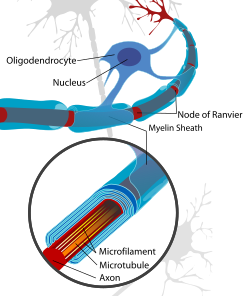 Oligodendrocytes are glial cells that form myelin around the axons of neurons.The brain is composed of two broad classes of cells: neurons and glia.[44] These two types are equally numerous in the brain as a whole, although glial cells outnumber neurons roughly 4 to 1 in the cerebral cortex.[45] Glia come in several types, which perform a number of critical functions, including structural support, metabolic support, insulation, and guidance of development.
The property that makes neurons so important is that, unlike glia, they are capable of sending signals to each other over long distances.[46] They send these signals by means of an axon, a thin protoplasmic fiber that extends from the cell body and projects, usually with numerous branches, to other areas, sometimes nearby, sometimes in distant parts of the brain or body. The extent of an axon can be extraordinary: to take an example, if a pyramidal cell of the neocortex were magnified so that its cell body became the size of a human, its axon, equally magnified, would become a cable a few centimeters in diameter, extending farther than a kilometer. These axons transmit signals in the form of electrochemical pulses called action potentials, lasting less than a thousandth of a second and traveling along the axon at speeds of 1–100 meters per second. Some neurons emit action potentials constantly, at rates of 10–100 per second, usually in irregular temporal patterns; other neurons are quiet most of the time, but occasionally emit a burst of action potentials.
 Neurons often have extensive networks of dendrites, which receive synaptic connections. Shown here is photomicrograph of a pyramidal neuron from the human hippocampus, stained by the Golgi method.Axons transmit signals to other neurons, or to non-neuronal cells, by means of specialized junctions called synapses.[47] A single axon may make as many as several thousand synaptic connections. When an action potential, traveling along an axon, arrives at a synapse, it causes a chemical called a neurotransmitter to be released. The neurotransmitter binds to receptor molecules in the membrane of the target cell. Some types of neuronal receptors are excitatory, meaning that they increase the rate of action potentials in the target cell; other receptors are inhibitory, meaning that they decrease the rate of action potentials; others have complex modulatory effects.
Axons actually fill most of the space in the brain.[48] Often large groups of them are bundled together in what are called nerve fiber tracts. Many axons are wrapped in thick sheaths of a fatty substance called myelin, which serves to greatly increase the speed of action potential propagation. Myelin is white, so parts of the brain filled exclusively with nerve fibers appear as white matter, in contrast to the gray matter that marks areas with high densities of neuron cell bodies. The total length of myelinated axons in an average adult human brain is well over 100,000 kilometres (62,000 mi) .[49]
 This diagram depicts the main subdivisions of the embryonic vertebrate brain. These regions will later differentiate into forebrain, midbrain and hindbrain structures.The brain does not simply grow; rather, it develops in an intricately orchestrated sequence of stages.[50] Many neurons are created in special zones that contain stem cells, and then migrate through the tissue to reach their ultimate locations.[51] In the cortex, for example, the first stage of development is the formation of a "scaffold" by a special group of glial cells, called radial glia, which send fibers vertically across the cortex. New cortical neurons are created at the bottom of the cortex, and then "climb" along the radial fibers until they reach the layers they are destined to occupy in the adult.
In vertebrates, the early stages of neural development are similar for all species.[50] As the embryo transforms from a round blob of cells into a wormlike structure, a narrow strip of ectoderm running along the midline of the back is induced to become the neural plate, the precursor of the nervous system. The neural plate invaginates to form the neural groove, and then the folds that line the groove merge to enclose the neural tube, a hollow cord of cells with a fluid-filled ventricle at the center. At the front end, the ventricles and cord swell to form three vesicles that are the precursors of the forebrain, midbrain, and hindbrain.[50] At the next stage, the forebrain splits into two vesicles called the telencephalon (which will contain the cerebral cortex, basal ganglia, and related structures) and the diencephalon (which will contain the thalamus and hypothalamus). At about the same time, the hindbrain splits into the metencephalon (which will contain the cerebellum and pons) and the myelencephalon (which will contain the medulla oblongata). Each of these areas contains proliferative zones at which neurons and glia cells are generated; the resulting cells then migrate, sometimes for long distances, to their final positions.
Once a neuron is in place, it begins to extend dendrites and an axon into the area around it.[52] Axons, because they commonly extend a great distance from the cell body and need to make contact with specific targets, grow in a particularly complex way. The tip of a growing axon consists of a blob of protoplasm called a "growth cone", studded with chemical receptors. These receptors sense the local environment, causing the growth cone to be attracted or repelled by various cellular elements, and thus to be pulled in a particular direction at each point along its path. The result of this pathfinding process is that the growth cone navigates through the brain until it reaches its destination area, where other chemical cues cause it to begin generating synapses. Taking the entire brain into account, many thousands of genes give rise to proteins that influence axonal pathfinding.
The synaptic network that finally emerges is only partly determined by genes, though. In many parts of the brain, axons initially "overgrow", and then are "pruned" by mechanisms that depend on neural activity.[53] In the projection from the eye to the midbrain, for example, the structure in the adult contains a very precise mapping, connecting each point on the surface of the retina to a corresponding point in a midbrain layer. In the first stages of development, each axon from the retina is guided to the right general vicinity in the midbrain by chemical cues, but then branches very profusely and makes initial contact with a wide swath of midbrain neurons. The retina, before birth, contains special mechanisms that cause it to generate waves of activity that originate spontaneously at some point and then propagate slowly across the retinal layer.[54] These waves are useful because they cause neighboring neurons to be active at the same time: that is, they produce a neural activity pattern that contains information about the spatial arrangement of the neurons. This information is exploited in the midbrain by a mechanism that causes synapses to weaken, and eventually vanish, if activity in an axon is not followed by activity of the target cell. The result of this sophisticated process is a gradual tuning and tightening of the map, leaving it finally in its precise adult form.
Similar things happen in other brain areas: an initial synaptic matrix is generated as a result of genetically determined chemical guidance, but then gradually refined by activity-dependent mechanisms, partly driven by internal dynamics, partly by external sensory inputs. In some cases, as with the retina-midbrain system, activity patterns depend on mechanisms that operate only in the developing brain, and apparently exist solely for the purpose of guiding development.[54]
In humans and many other mammals, new neurons are created mainly before birth, and the infant brain actually contains substantially more neurons than the adult brain.[55] There are, however, a few areas where new neurons continue to be generated throughout life. The two areas for which this is well established are the olfactory bulb, which is involved in the sense of smell, and the dentate gyrus of the hippocampus, where there is evidence that the new neurons play a role in storing newly acquired memories. With these exceptions, however, the set of neurons that is present in early childhood is the set that is present for life. Glial cells are different, however; as with most types of cells in the body, these are generated throughout the lifespan.
Although the pool of neurons is largely in place by birth, the axonal connections continue to develop for a long time afterward. In humans, full myelination is not completed until adolescence.[56]
There has long been debate about whether the qualities of mind, personality, and intelligence can mainly be attributed to heredity or to upbringing; the nature versus nurture debate.[57] This is not just a philosophical question: it has great practical relevance to parents and educators. Although many details remain to be settled, neuroscience clearly shows that both factors are essential. Genes determine the general form of the brain, and genes determine how the brain reacts to experience. Experience, however, is required to refine the matrix of synaptic connections. In some respects it is mainly a matter of presence or absence of experience during critical periods of development.[58] In other respects, the quantity and quality of experience may be more relevant: for example, there is substantial evidence that animals raised in enriched environments have thicker cortices, indicating a higher density of synaptic connections, than animals whose levels of stimulation are restricted.[59]
Nevertheless, the great majority of psychoactive drugs exert their effects by altering neurotransmitter systems not directly involving glutamatergic or GABAergic transmission.[63] Drugs such as caffeine, nicotine, heroin, cocaine, Prozac, Thorazine, etc., act on other neurotransmitters. Many of these other transmitters come from neurons that are localized in particular parts of the brain. Serotonin, for example—the primary target of antidepressant drugs and many dietary aids—comes exclusively from a small brainstem area called the Raphe nuclei. Norepinephrine, which is involved in arousal, comes exclusively from a nearby small area called the locus ceruleus. Histamine, as a neurotransmitter, comes from a tiny part of the hypothalamus called the tuberomammilary nucleus (histamine also has non-CNS functions, but the neurotransmitter function is what causes antihistamines to have sedative effects). Other neurotransmitters such as acetylcholine and dopamine have multiple sources in the brain, but are not as ubiquitously distributed as glutamate and GABA.
Each sensory system begins with specialized "sensory receptor" cells. These are neurons, but unlike most neurons, they are not controlled by synaptic input from other neurons: instead they are activated by membrane-bound receptors that are sensitive to some physical modality, such as light, temperature, or physical stretching. The axons of sensory receptor cells travel into the spinal cord or brain. For the sense of hearing, the receptors are located in the inner ear, on the cochlea, and are activated by vibration.[65]
For most senses, there is a "primary nucleus" or set of nuclei, located in the brainstem, that gathers signals from the sensory receptor cells. For the sense of hearing, these are the cochlear nuclei.[65] In many cases, there are secondary subcortical areas that extract special information of some sort. For the sense of hearing, the superior olivary area and inferior colliculus are involved in comparing the signals from the two ears to extract information about the direction of the sound source, among other functions.[65] Each sensory system also has a special part of the thalamus dedicated to it, which serves as a relay to the cortex. For the sense of hearing, this is the medial geniculate nucleus.[65]
For each sensory system, there is a "primary" cortical area that receives direct input from the thalamic relay area. For the auditory system this is the primary auditory cortex, located in the upper part of the temporal lobe.[65] There are also usually a set of "higher level" cortical sensory areas, which analyze the sensory input in specific ways. For the auditory system, there are areas that analyze sound quality, rhythm, and temporal patterns of change, among other features.[65] Finally, there are multimodal areas that combine inputs from different sensory modalities, for example auditory and visual. At this point, the signals have reached parts of the brain that are best described as integrative rather than specifically sensory.[65]
All of these rules have exceptions. For example, the sense of touch (which is actually a set of at least half-a-dozen distinct mechanical senses), the sensory inputs terminate mainly in the spinal cord, on neurons that then project to the brainstem.[66] For the sense of smell, there is no relay in the thalamus; instead the signals go directly from the primary brain area—the olfactory bulb—to the cortex.[67]
The brain contains a number of areas that project directly to the spinal cord.[71] At the lowest level are motor areas in the medulla and pons. At a higher level are areas in the midbrain, such as the red nucleus, which is responsible for coordinating movements of the arms and legs. At a higher level yet is the primary motor cortex, a strip of tissue located at the posterior edge of the frontal lobe. The primary motor cortex sends projections to the subcortical motor areas, but also sends a massive projection directly to the spinal cord, via the so-called pyramidal tract. This direct corticospinal projection allows for precise voluntary control of the fine details of movements. Other "secondary" motor-related brain areas do not project directly to the spinal cord, but instead act on the cortical or subcortical primary motor areas. Among the most important secondary areas are the premotor cortex, basal ganglia, and cerebellum:
A key component of the arousal system is the suprachiasmatic nucleus (SCN), a tiny part of the hypothalamus located directly above the point at which the optic nerves from the two eyes cross.[77] The SCN contains the body's central biological clock. Neurons there show activity levels that rise and fall with a period of about 24 hours, circadian rhythms: these activity fluctuations are driven by rhythmic changes in expression of a set of "clock genes". The SCN continues to keep time even if it is excised from the brain and placed in a dish of warm nutrient solution, but it ordinarily receives input from the optic nerves, through the retinohypothalamic tract (RHT), that allow daily light-dark cycles to calibrate the clock.
The SCN projects to a set of areas in the hypothalamus, brainstem, and midbrain that are involved in implementing sleep-wake cycles. An important component of the system is the so-called reticular formation, a group of neuron-clusters scattered diffusely through the core of the lower brain.[76] Reticular neurons send signals to the thalamus, which in turn sends activity-level-controlling signals to every part of the cortex. Damage to the reticular formation can produce a permanent state of coma.
Sleep involves great changes in brain activity.[78] Until the 1950s it was generally believed that the brain essentially shuts off during sleep,[79] but this is now known to be far from true: activity continues, but patterns become very different. In fact, there are two types of sleep, REM sleep (with dreaming) and NREM (non-REM, usually without dreaming) sleep, which repeat in slightly varying patterns throughout a sleep episode. Three broad types of distinct brain activity patterns can be measured: REM, light NREM and deep NREM. During deep NREM sleep, also called slow wave sleep, activity in the cortex takes the form of large synchronized waves, where in the waking state it is noisy and desynchronized. Levels of the neurotransmitters norepinephrine and serotonin drop during slow wave sleep, and fall almost to zero during REM sleep; levels of acetylcholine show the reverse pattern.
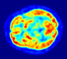 PET Image of the human brain showing energy consumptionAlthough the human brain represents only 2% of the body weight, it receives 15% of the cardiac output, 20% of total body oxygen consumption, and 25% of total body glucose utilization.[80] The need to limit body weight in order, for example, to fly, has led to selection for a reduction of brain size in some species, such as bats.[81] The brain mostly utilizes glucose for energy, and deprivation of glucose, as can happen in hypoglycemia, can result in loss of consciousness. The energy consumption of the brain does not vary greatly over time, but active regions of the cortex consume somewhat more energy than inactive regions: this fact forms the basis for the functional brain imaging methods PET and fMRI.[82] These are nuclear medicine imaging techniques which produce a three-dimensional image of metabolic activity.
The mind-body problem is one of the central issues in the history of philosophy,[85] which asks us to consider if the brain and the mind are identical, partially distinct, or related in some unknown way. There are three major schools of thought concerning the answer: dualism, materialism, and idealism. Dualism holds that the mind exists independently of the brain;[86] materialism holds that mental phenomena are identical to neuronal phenomena;[87] and idealism holds that only mental phenomena exist.[87]
In addition to the philosophical questions, the relationship between mind and brain involves a high number of scientific questions, including understanding the relationship between mental activity and brain activity, the exact mechanisms by which drugs influence cognition, and the neural correlates of consciousness.
Through most of history many philosophers found it inconceivable that cognition could be implemented by a physical substance such as brain tissue (that is neurons and synapses).[88] Philosophers such as Patricia Churchland posit that the drug-mind interaction is indicative of an intimate connection between the brain and the mind, not that the two are the same entity.[89] Descartes, who thought extensively about mind-brain relationships, found it possible to explain reflexes and other simple behaviors in mechanistic terms, although he did not believe that complex thought, and language in particular, could be explained by reference to the physical brain alone.[90]
The oldest method of studying the brain is anatomical, and until the middle of the 20th century, much of the progress in neuroscience came from the development of better stains and better microscopes—the neuroanatomist Floyd Bloom famously quipped that "the gain in brain is mainly in the stain."[93] Neuroanatomists study the large-scale structure of the brain as well as the microscopic structure of neurons and their components, especially synapses. Among other tools, they employ a plethora of stains that reveal neural structure, chemistry, and connectivity. In recent years, the development of immunostaining techniques has allowed staining of neurons that express specific sets of genes. Also, functional neuroanatomy uses medical imaging techniques to correlate variations in human brain structure with differences in cognition or behavior.
Neurophysiologists study the chemical, pharmacological, and electrical properties of the brain: their primary tools are drugs and recording devices. Many thousands of experimentally developed drugs affect the nervous system, some in highly specific ways. Recordings of brain activity can be made using electrodes, either glued to the skull as in EEG studies, or implanted inside the brains of animals for extracellular recordings, which can detect action potentials generated by individual neurons.[94] Because the brain does not contain pain receptors, it is possible using these techniques to record from animals that are awake and behaving without causing distress. The same techniques have occasionally been used to study brain activity in human patients suffering from intractable epilepsy, in cases where there was a medical necessity to implant electrodes in order to localize the brain area responsible for seizures.[95] It is also possible to study brain activity noninvasively in humans using functional imaging techniques such as MRI—this field has expanded enormously over the past two decades.
A different approach to brain function is to examine the consequences of damage to specific brain areas. Even though it is protected by the skull and meninges, surrounded by cerebrospinal fluid, and isolated from the bloodstream by the blood-brain barrier, the delicate nature of the brain makes it vulnerable to numerous diseases and several types of damage. In humans, the effects of strokes and other types of brain damage have been a key source of information about brain function.[96] Because there is no ability to experimentally control the nature of the damage, however, this information is often difficult to interpret. In animal studies, most commonly involving rats, it is possible to use electrodes or locally injected chemicals to produce precise patterns of damage and then examine the consequences for behavior.
Computational neuroscience encompasses two approaches: first, the use of computers to study the brain; second, the study of how brains perform computation.[97] On one hand, it is possible to write a computer program to simulate the operation of a group of neurons by making use of systems of equations that describe their electrochemical activity; such simulations are known as biologically realistic neural networks. On the other hand, it is possible to study algorithms for neural computation by simulating, or mathematically analyzing, the operations of simplified "units" that have some of the properties of neurons but abstract out much of their biological complexity. The computational functions of the brain are studied both by neuroscientists and computer scientists.
Recent years have seen the first applications of genetic engineering techniques to the study of the brain.[98] The most common subjects are mice, because the technical tools are more advanced for this species than for any other. It is now possible with relative ease to "knock out" or mutate a wide variety of genes, and then examine the effects on brain function. More sophisticated approaches are also beginning to be used: for example, using the Cre-Lox recombination method it is possible to activate or inactivate genes in specific parts of the brain, at specific times.
 Drawing by Santiago Ramon y Cajal of two types of Golgi-stained neurons from the cerebellum of a pigeonEach major advance in understanding has followed more or less directly from the development of a new method of investigation. Until the early years of the 20th century, the most important advances were derived from new stains.[102] Particularly critical was the invention of the Golgi stain, which (when correctly used) stains only a small, and apparently random, fraction of neurons, but stains them in their entirety, including cell body, dendrites, and axon. Without such a stain, brain tissue under a microscope appears as an impenetrable tangle of protoplasmic fibers, in which it is impossible to determine any structure. In the hands of Camillo Golgi, and especially of the Spanish neuroanatomist Santiago Ramon y Cajal, the new stain revealed hundreds of distinct types of neurons, each with its own unique dendritic structure and pattern of connectivity.
In the 20th century, progress in electronics enabled investigation of the electrical properties of nerve cells, culminating in the work by Alan Hodgkin, Andrew Huxley, and others on the biophysics of the action potential, and the work of Bernard Katz and others on the electrochemistry of the synapse.[103] The earliest studies used special preparations, such as the "fast escape response" system of the squid, which involves a giant axon as thick as a pencil lead, and giant synapses connecting to this axon. Steady improvements in electrodes and electronics allowed ever finer levels of resolution. These studies complemented the anatomical picture with a conception of the brain as a dynamic entity. Reflecting the new understanding, in 1942 Charles Sherrington visualized the workings of the brain in action in somewhat breathless terms:
The great topmost sheet of the mass, that where hardly a light had twinkled or moved, becomes now a sparkling field of rhythmic flashing points with trains of traveling sparks hurrying hither and thither. ... It is as if the Milky Way entered upon some cosmic dance. Swiftly the head mass becomes an enchanted loom where millions of flashing shuttles weave a dissolving pattern, always a meaningful pattern though never an abiding one; a shifting harmony of subpatterns.
Brains can be extremely complex. The cerebral cortex of the human brain contains roughly 15–33 billion neurons, perhaps more, depending on gender and age,[2] linked with up to 10,000 synaptic connections each. Each cubic millimeter of cerebral cortex contains roughly one billion synapses.[3] These neurons communicate with one another by means of long protoplasmic fibers called axons, which carry trains of signal pulses called action potentials to distant parts of the brain or body and target them to specific recipient cells.
The brain controls the other organ systems of the body, either by activating muscles or by causing secretion of chemicals such as hormones and neurotransmitters. This centralized control allows rapid and coordinated responses to changes in the environment. Some basic types of responsiveness are possible without a brain: even single-celled organisms may be capable of extracting information from the environment and acting in response to it.[4] Sponges, which lack a central nervous system, are capable of coordinated body contractions and even locomotion.[5] In vertebrates, the spinal cord by itself contains neural circuitry capable of generating reflex responses as well as simple motor patterns such as swimming or walking.[6] However, sophisticated control of behavior on the basis of complex sensory input requires the information-integrating capabilities of a centralized brain.
Despite rapid scientific progress, much about how brains work remains a mystery. The operations of individual neurons and synapses are now understood in considerable detail, but the way they cooperate in ensembles of thousands or millions has been very difficult to decipher. Methods of observation such as EEG recording and functional brain imaging tell us that brain operations are highly organized, while single unit recording can resolve the activity of single neurons, but how individual cells give rise to complex operations is unknown.[7]

Contents
[hide]- 1 Macroscopic structure
- 2 Microscopic structure
- 3 Development
- 4 Functions
- 5 Brain and mind
- 6 Research
- 7 History
- 8 See also
- 9 Notes
- 10 References
- 11 Further reading
- 12 External links
Macroscopic structure
The brain is the most complex biological structure known,[8] and comparing the brains of different species on the basis of appearance is often difficult. Nevertheless, there are common principles of brain architecture that apply across a wide range of species. These are revealed mainly by three approaches. The evolutionary approach compares brain structures of different species, and using the principle that features found in all branches that have descended from a given ancient form were probably present in the common ancestor as well. The developmental approach examines how the form of the brain changes during the progression from embryonic to adult stages. The genetic approach analyzes gene expression in various parts of the brain across a range of species. Each approach complements and informs the other two.Bilateria

All bilateria are thought to have descended from a common ancestor that appeared early in the Cambrian period, 550–600 million years ago.[9] This ancestor had the shape of a simple tube worm with a segmented body, and at an abstract level, that worm-shape continues to be reflected in the body and nervous system plans of all modern bilateria, including humans.[10] The fundamental bilateral body form is a tube with a hollow gut cavity running from mouth to anus, and a nerve cord with an enlargement (a "ganglion") for each body segment, with an especially large ganglion at the front, called the "brain".
Invertebrates
For invertebrates (e.g., insects, molluscs, worms, etc.) the components of the brain differ so greatly from the vertebrate pattern that it is hard to make meaningful comparisons except on the basis of genetics. Two groups of invertebrates have notably complex brains: arthropods (insects, crustaceans, arachnids, and others), and cephalopods (octopuses, squids, and similar molluscs).[11] The brains of arthropods and cephalopods arise from twin parallel nerve cords that extend through the body of the animal. Arthropods have a central brain with three divisions and large optical lobes behind each eye for visual processing.[11] Cephalopods have the largest brains of any invertebrates. The brain of the octopus in particular is highly developed, comparable in complexity to the brains of some vertebrates.
There are a few invertebrates whose brains have been studied intensively. The large sea slug Aplysia was chosen by Nobel Prize-winning neurophysiologist Eric Kandel, because of the simplicity and accessibility of its nervous system, as a model for studying the cellular basis of learning and memory, and subjected to hundreds of experiments.[12] The most thoroughly studied invertebrate brains, however, belong to the fruit fly Drosophila and the tiny roundworm Caenorhabditis elegans (C. elegans).
Because of the large array of techniques available for studying their genetics, fruit flies have been a natural subject for studying the role of genes in brain development.[13] Remarkably, many aspects of Drosophila neurogenetics have turned out to be relevant to humans. The first biological clock genes, for example, were identified by examining Drosophila mutants that showed disrupted daily activity cycles.[14] A search in the genomes of vertebrates turned up a set of analogous genes, which were found to play similar roles in the mouse biological clock—and therefore almost certainly in the human biological clock as well.[15]
Like Drosophila, the nematode worm C. elegans has been studied largely because of its importance in genetics.[16] In the early 1970s, Sydney Brenner chose it as a model system for studying the way that genes control development. One of the advantages of working with this worm is that the body plan is very stereotyped: the nervous system of the hermaphrodite morph contains exactly 302 neurons, always in the same places, making identical synaptic connections in every worm.[17] In a heroic project, Brenner's team sliced worms into thousands of ultrathin sections and photographed every section under an electron microscope, then visually matched fibers from section to section, in order to map out every neuron and synapse in the entire body.[18] Nothing approaching this level of detail is available for any other organism, and the information has been used to enable a multitude of studies that would not have been possible without it.
Vertebrates

The first vertebrates appeared over 500 million years ago (mya), during the Cambrian period, and may have somewhat resembled the modern hagfish in form.[21] Sharks appeared about 450 mya, amphibians about 400 mya, reptiles about 350 mya, and mammals about 200 mya. No modern species should be described as more "primitive" than others, since all have an equally long evolutionary history, but the brains of modern hagfishes, lampreys, sharks, amphibians, reptiles, and mammals show a gradient of size and complexity that roughly follows the evolutionary sequence.[22] All of these brains contain the same set of basic anatomical components, but many are rudimentary in hagfishes, whereas in mammals the foremost parts are greatly elaborated and expanded.
All vertebrate brains share a common underlying form, which can most easily be appreciated by examining how they develop.[23] The first appearance of the nervous system is as a thin strip of tissue running along the back of the embryo. This strip thickens and then folds up to form a hollow tube. The front end of the tube develops into the brain. In its earliest form, the brain appears as three swellings, which eventually become the forebrain, midbrain, and hindbrain. In many classes of vertebrates these three parts remain similar in size in the adult, but in mammals the forebrain becomes much larger than the other parts, and the midbrain quite small.
The relationship between brain size, body size and other variables has been studied across a wide range of vertebrate species. Brain size increases with body size but not proportionally. Averaging across all orders of mammals, it follows a power law, with an exponent of about 0.75.[24] This formula applies to the average brain of mammals but each family departs from it, reflecting their sophistication of behavior.[25] For example, primates have brains 5 to 10 times as large as the formula predicts. Predators tend to have larger brains. When the mammalian brain increases in size, not all parts increase at the same rate. The larger the brain of a species, the greater the fraction taken up by the cortex.[26]
Vertebrate brain regions
Neuroanatomists usually consider the brain to consist of six main regions: the telencephalon (cerebral hemispheres), diencephalon (thalamus and hypothalamus), mesencephalon (midbrain), cerebellum, pons, and medulla oblongata.[27] Each of these areas in turn has a complex internal structure. Some areas, such as the cortex and cerebellum, consist of layers, folded or convoluted to fit within the available space. Other areas consist of clusters of many small nuclei. If fine distinctions are made on the basis of neural structure, chemistry, and connectivity, thousands of distinguishable areas can be identified within the vertebrate brain.Some branches of vertebrate evolution have led to substantial changes in brain shape, especially in the forebrain. The brain of a shark shows the basic components in a straightforward way, but in teleost fishes (the great majority of modern species), the forebrain has become "everted", like a sock turned inside out. In birds, also, there are major changes in shape.[28] One of the main structures in the avian forebrain, the dorsal ventricular ridge, was long thought to correspond to the basal ganglia of mammals, but is now thought to be more closely related to the neocortex.[29]

- The medulla, along with the spinal cord, contains many small nuclei involved in a wide variety of sensory and motor functions.[30]
- The hypothalamus is a small region at the base of the forebrain, whose complexity and importance belies its size. It is composed of numerous small nuclei, each with distinct connections and distinct neurochemistry. The hypothalamus is the central control station for sleep/wake cycles, control of eating and drinking, control of hormone release, and many other critical biological functions.[31]
- Like the hypothalamus, the thalamus is a collection of nuclei with diverse functions. Some of them are involved in relaying information to and from the cerebral hemispheres. Others are involved in motivation. The subthalamic area (zona incerta) seems to contain action-generating systems for several types of "consummatory" behaviors, including eating, drinking, defecation, and copulation.[32]
- The cerebellum modulates the outputs of other brain systems to make them more precise. Removal of the cerebellum does not prevent an animal from doing anything in particular, but it makes actions hesitant and clumsy. This precision is not built-in, but learned by trial and error. Learning how to ride a bicycle is an example of a type of neural plasticity that may take place largely within the cerebellum.[33]
- The tectum, often called "optic tectum", allows actions to be directed toward points in space. In mammals it is called the "superior colliculus", and its best studied function is to direct eye movements. It also directs reaching movements, though. It gets strong visual inputs, but also inputs from other senses that are useful in directing actions, such as auditory input in owls, input from the thermosensitive pit organs in snakes, etc. In some fishes, such as lampreys, it is the largest part of the brain.[34]
- The pallium is a layer of gray matter that lies on the surface of the forebrain. In reptiles and mammals it is called cortex instead. The pallium is involved in multiple functions, including olfaction and spatial memory. In mammals, where it comes to dominate the brain, it subsumes functions from many subcortical areas.[35]
- The hippocampus, strictly speaking, is found only in mammals. However, the area it derives from, the medial pallium, has counterparts in all vertebrates. There is evidence that this part of the brain is involved in spatial memory and navigation in fishes, birds, reptiles, and mammals.[36]
- The basal ganglia are a group of interconnected structures in the forebrain, of which our understanding has increased enormously over the last few years. The primary function of the basal ganglia seems to be action selection. They send inhibitory signals to all parts of the brain that can generate actions, and in the right circumstances can release the inhibition, so that the action-generating systems are able to execute their actions. Rewards and punishments exert their most important neural effects within the basal ganglia.[37]
- The olfactory bulb is a special structure that processes olfactory sensory signals, and sends its output to the olfactory part of the pallium. It is a major brain component in many vertebrates, but much reduced in primates.[38]
Mammals
The cerebral cortex is the part of the brain that most strongly distinguishes mammals from other vertebrates, primates from other mammals, and humans from other primates. The hindbrain and midbrain of mammals are generally similar to those of other vertebrates, but dramatic differences appear in the forebrain, which is not only greatly enlarged, but also altered in structure.[39] In non-mammalian vertebrates, the surface of the cerebrum is lined with a comparatively simple layered structure called the pallium.[40] In mammals, the pallium evolves into a complex 6-layered structure called neocortex or isocortex. In primates, the neocortex is greatly enlarged, especially the part called the frontal lobes. In humans, this enlargement of the frontal lobes is taken to an extreme, and other parts of the cortex also become quite large and complex. The hippocampus of mammals also has a distinctive structure.
Primates
See also: Human brain
The brains of humans and other primates contain the same structures as the brains of other mammals, but are considerably larger in proportion to body size.[26] Most of the enlargement comes from a massive expansion of the cortex, focusing especially on the parts serving vision and forethought.[42] The visual processing network of primates is very complex, including at least 30 distinguishable areas, with a bewildering web of interconnections. Taking all of these together, visual processing makes use of more than half of the primate neocortex.[43] The other part of the brain that is greatly enlarged is the prefrontal cortex, whose functions are difficult to summarize succinctly, but relate to planning, working memory, motivation, attention, and executive control.Microscopic structure

The property that makes neurons so important is that, unlike glia, they are capable of sending signals to each other over long distances.[46] They send these signals by means of an axon, a thin protoplasmic fiber that extends from the cell body and projects, usually with numerous branches, to other areas, sometimes nearby, sometimes in distant parts of the brain or body. The extent of an axon can be extraordinary: to take an example, if a pyramidal cell of the neocortex were magnified so that its cell body became the size of a human, its axon, equally magnified, would become a cable a few centimeters in diameter, extending farther than a kilometer. These axons transmit signals in the form of electrochemical pulses called action potentials, lasting less than a thousandth of a second and traveling along the axon at speeds of 1–100 meters per second. Some neurons emit action potentials constantly, at rates of 10–100 per second, usually in irregular temporal patterns; other neurons are quiet most of the time, but occasionally emit a burst of action potentials.

Axons actually fill most of the space in the brain.[48] Often large groups of them are bundled together in what are called nerve fiber tracts. Many axons are wrapped in thick sheaths of a fatty substance called myelin, which serves to greatly increase the speed of action potential propagation. Myelin is white, so parts of the brain filled exclusively with nerve fibers appear as white matter, in contrast to the gray matter that marks areas with high densities of neuron cell bodies. The total length of myelinated axons in an average adult human brain is well over 100,000 kilometres (62,000 mi) .[49]
Development
Main article: Neural development

In vertebrates, the early stages of neural development are similar for all species.[50] As the embryo transforms from a round blob of cells into a wormlike structure, a narrow strip of ectoderm running along the midline of the back is induced to become the neural plate, the precursor of the nervous system. The neural plate invaginates to form the neural groove, and then the folds that line the groove merge to enclose the neural tube, a hollow cord of cells with a fluid-filled ventricle at the center. At the front end, the ventricles and cord swell to form three vesicles that are the precursors of the forebrain, midbrain, and hindbrain.[50] At the next stage, the forebrain splits into two vesicles called the telencephalon (which will contain the cerebral cortex, basal ganglia, and related structures) and the diencephalon (which will contain the thalamus and hypothalamus). At about the same time, the hindbrain splits into the metencephalon (which will contain the cerebellum and pons) and the myelencephalon (which will contain the medulla oblongata). Each of these areas contains proliferative zones at which neurons and glia cells are generated; the resulting cells then migrate, sometimes for long distances, to their final positions.
Once a neuron is in place, it begins to extend dendrites and an axon into the area around it.[52] Axons, because they commonly extend a great distance from the cell body and need to make contact with specific targets, grow in a particularly complex way. The tip of a growing axon consists of a blob of protoplasm called a "growth cone", studded with chemical receptors. These receptors sense the local environment, causing the growth cone to be attracted or repelled by various cellular elements, and thus to be pulled in a particular direction at each point along its path. The result of this pathfinding process is that the growth cone navigates through the brain until it reaches its destination area, where other chemical cues cause it to begin generating synapses. Taking the entire brain into account, many thousands of genes give rise to proteins that influence axonal pathfinding.
The synaptic network that finally emerges is only partly determined by genes, though. In many parts of the brain, axons initially "overgrow", and then are "pruned" by mechanisms that depend on neural activity.[53] In the projection from the eye to the midbrain, for example, the structure in the adult contains a very precise mapping, connecting each point on the surface of the retina to a corresponding point in a midbrain layer. In the first stages of development, each axon from the retina is guided to the right general vicinity in the midbrain by chemical cues, but then branches very profusely and makes initial contact with a wide swath of midbrain neurons. The retina, before birth, contains special mechanisms that cause it to generate waves of activity that originate spontaneously at some point and then propagate slowly across the retinal layer.[54] These waves are useful because they cause neighboring neurons to be active at the same time: that is, they produce a neural activity pattern that contains information about the spatial arrangement of the neurons. This information is exploited in the midbrain by a mechanism that causes synapses to weaken, and eventually vanish, if activity in an axon is not followed by activity of the target cell. The result of this sophisticated process is a gradual tuning and tightening of the map, leaving it finally in its precise adult form.
Similar things happen in other brain areas: an initial synaptic matrix is generated as a result of genetically determined chemical guidance, but then gradually refined by activity-dependent mechanisms, partly driven by internal dynamics, partly by external sensory inputs. In some cases, as with the retina-midbrain system, activity patterns depend on mechanisms that operate only in the developing brain, and apparently exist solely for the purpose of guiding development.[54]
In humans and many other mammals, new neurons are created mainly before birth, and the infant brain actually contains substantially more neurons than the adult brain.[55] There are, however, a few areas where new neurons continue to be generated throughout life. The two areas for which this is well established are the olfactory bulb, which is involved in the sense of smell, and the dentate gyrus of the hippocampus, where there is evidence that the new neurons play a role in storing newly acquired memories. With these exceptions, however, the set of neurons that is present in early childhood is the set that is present for life. Glial cells are different, however; as with most types of cells in the body, these are generated throughout the lifespan.
Although the pool of neurons is largely in place by birth, the axonal connections continue to develop for a long time afterward. In humans, full myelination is not completed until adolescence.[56]
There has long been debate about whether the qualities of mind, personality, and intelligence can mainly be attributed to heredity or to upbringing; the nature versus nurture debate.[57] This is not just a philosophical question: it has great practical relevance to parents and educators. Although many details remain to be settled, neuroscience clearly shows that both factors are essential. Genes determine the general form of the brain, and genes determine how the brain reacts to experience. Experience, however, is required to refine the matrix of synaptic connections. In some respects it is mainly a matter of presence or absence of experience during critical periods of development.[58] In other respects, the quantity and quality of experience may be more relevant: for example, there is substantial evidence that animals raised in enriched environments have thicker cortices, indicating a higher density of synaptic connections, than animals whose levels of stimulation are restricted.[59]
Functions
From an organismic perspective, the primary function of a brain is to control the actions of an animal.[60] To do this, it extracts enough relevant information from sense organs to refine actions. Sensory signals may stimulate an immediate response as when the olfactory system of a deer detects the odor of a wolf; they may modulate an ongoing pattern of activity as in the effect of light-dark cycles on an organism's sleep-wake behavior; or their information may be stored in case of future relevance. The brain manages its complex task by orchestrating functional subsystems, which can be categorized in a number of ways: anatomically, chemically, and functionally.Functional subsystems
The most straightforward way to categorize the parts of the brain is anatomically, but there are also several ways to subdivide it functionally. One of the most important of these is on the basis of the chemical neurotransmitters used by neurons to communicate with each other. Another is in terms of the way a brain area contributes to information processing: sensory areas bring information into the brain and reformat it; motor signals send information out of the brain to control muscles and glands; arousal systems modulate the activity of the brain according to time of day and other factors.Neurotransmitter systems
Main article: Neurotransmitter systems
With few exceptions, each neuron in the brain consistently releases the same chemical neurotransmitter, or combination of neurotransmitters, at all of the synaptic connections it makes with other neurons; this rule is known as Dale's principle.[61] Thus, a neuron can be characterized by the neurotransmitters it releases. The two neurotransmitters that appear most frequently are glutamate, which is almost always excitatory, and gamma-aminobutyric acid (GABA), which is almost always inhibitory. Neurons using these transmitters can be found in nearly every part of the brain, making up a large percentage of the brain's pool of synapses.[62]Nevertheless, the great majority of psychoactive drugs exert their effects by altering neurotransmitter systems not directly involving glutamatergic or GABAergic transmission.[63] Drugs such as caffeine, nicotine, heroin, cocaine, Prozac, Thorazine, etc., act on other neurotransmitters. Many of these other transmitters come from neurons that are localized in particular parts of the brain. Serotonin, for example—the primary target of antidepressant drugs and many dietary aids—comes exclusively from a small brainstem area called the Raphe nuclei. Norepinephrine, which is involved in arousal, comes exclusively from a nearby small area called the locus ceruleus. Histamine, as a neurotransmitter, comes from a tiny part of the hypothalamus called the tuberomammilary nucleus (histamine also has non-CNS functions, but the neurotransmitter function is what causes antihistamines to have sedative effects). Other neurotransmitters such as acetylcholine and dopamine have multiple sources in the brain, but are not as ubiquitously distributed as glutamate and GABA.
Sensory systems
Main article: Sensory system
One of the primary functions of a brain is to extract biologically relevant information from sensory inputs.[64] Even in the human brain, sensory processes go well beyond the classical five senses of sight, sound, taste, touch, and smell: our brains are provided with information about temperature, balance, limb position, and the chemical composition of the bloodstream, among other things. All of these modalities are detected by specialized sensors that project signals into the brain. In other animals, additional senses may be present, such as the infrared heat-sensors in the pit organs of snakes; or the "standard" senses may be used in nonstandard ways, as in the auditory "sonar" of bats. Here are a few principles that apply to most sensory systems, using the auditory system for specific examples.Each sensory system begins with specialized "sensory receptor" cells. These are neurons, but unlike most neurons, they are not controlled by synaptic input from other neurons: instead they are activated by membrane-bound receptors that are sensitive to some physical modality, such as light, temperature, or physical stretching. The axons of sensory receptor cells travel into the spinal cord or brain. For the sense of hearing, the receptors are located in the inner ear, on the cochlea, and are activated by vibration.[65]
For most senses, there is a "primary nucleus" or set of nuclei, located in the brainstem, that gathers signals from the sensory receptor cells. For the sense of hearing, these are the cochlear nuclei.[65] In many cases, there are secondary subcortical areas that extract special information of some sort. For the sense of hearing, the superior olivary area and inferior colliculus are involved in comparing the signals from the two ears to extract information about the direction of the sound source, among other functions.[65] Each sensory system also has a special part of the thalamus dedicated to it, which serves as a relay to the cortex. For the sense of hearing, this is the medial geniculate nucleus.[65]
For each sensory system, there is a "primary" cortical area that receives direct input from the thalamic relay area. For the auditory system this is the primary auditory cortex, located in the upper part of the temporal lobe.[65] There are also usually a set of "higher level" cortical sensory areas, which analyze the sensory input in specific ways. For the auditory system, there are areas that analyze sound quality, rhythm, and temporal patterns of change, among other features.[65] Finally, there are multimodal areas that combine inputs from different sensory modalities, for example auditory and visual. At this point, the signals have reached parts of the brain that are best described as integrative rather than specifically sensory.[65]
All of these rules have exceptions. For example, the sense of touch (which is actually a set of at least half-a-dozen distinct mechanical senses), the sensory inputs terminate mainly in the spinal cord, on neurons that then project to the brainstem.[66] For the sense of smell, there is no relay in the thalamus; instead the signals go directly from the primary brain area—the olfactory bulb—to the cortex.[67]
Motor systems
Main article: Motor system
Motor systems are areas of the brain that are more or less directly involved in producing body movements, that is, in activating muscles. With the exception of the muscles that control the eye, all of the voluntary muscles[68] in the body are directly innervated by motor neurons in the spinal cord, which therefore are the final common path for the movement-generating system.[69] Spinal motor neurons are controlled both by neural circuits intrinsic to the spinal cord, and by inputs that descend from the brain. The intrinsic spinal circuits implement many reflex responses, and also contain pattern generators for rhythmic movements such as walking or swimming.[70] The descending connections from the brain allow for more sophisticated control.The brain contains a number of areas that project directly to the spinal cord.[71] At the lowest level are motor areas in the medulla and pons. At a higher level are areas in the midbrain, such as the red nucleus, which is responsible for coordinating movements of the arms and legs. At a higher level yet is the primary motor cortex, a strip of tissue located at the posterior edge of the frontal lobe. The primary motor cortex sends projections to the subcortical motor areas, but also sends a massive projection directly to the spinal cord, via the so-called pyramidal tract. This direct corticospinal projection allows for precise voluntary control of the fine details of movements. Other "secondary" motor-related brain areas do not project directly to the spinal cord, but instead act on the cortical or subcortical primary motor areas. Among the most important secondary areas are the premotor cortex, basal ganglia, and cerebellum:
- The premotor cortex (which is actually a large complex of areas) adjoins the primary motor cortex, and projects to it. Whereas elements of the primary motor cortex map to specific body areas, elements of the premotor cortex are often involved in coordinated movements of multiple body parts.[72]
- The basal ganglia are a set of structures in the base of the forebrain that project to many other motor-related areas.[73] Their function has been difficult to understand, but one of the most popular theories currently is that they play a key role in action selection.[74] Most of the time they restrain actions by sending constant inhibitory signals to action-generating systems, but in the right circumstances, they release this inhibition and therefore allow their targets to take control of behavior.
- The cerebellum is a very distinctive structure attached to the back of the brain.[33] It does not control or originate behaviors, but instead generates corrective signals to make movements more precise. People with cerebellar damage are not paralyzed in any way, but their body movements become erratic and uncoordinated.
Arousal systems
See also: Sleep
Perhaps the most obvious aspect of the behavior of any animal is the daily cycle between sleeping and waking. Arousal and alertness are also modulated on a finer time scale, though, by an extensive network of brain areas.[76]A key component of the arousal system is the suprachiasmatic nucleus (SCN), a tiny part of the hypothalamus located directly above the point at which the optic nerves from the two eyes cross.[77] The SCN contains the body's central biological clock. Neurons there show activity levels that rise and fall with a period of about 24 hours, circadian rhythms: these activity fluctuations are driven by rhythmic changes in expression of a set of "clock genes". The SCN continues to keep time even if it is excised from the brain and placed in a dish of warm nutrient solution, but it ordinarily receives input from the optic nerves, through the retinohypothalamic tract (RHT), that allow daily light-dark cycles to calibrate the clock.
The SCN projects to a set of areas in the hypothalamus, brainstem, and midbrain that are involved in implementing sleep-wake cycles. An important component of the system is the so-called reticular formation, a group of neuron-clusters scattered diffusely through the core of the lower brain.[76] Reticular neurons send signals to the thalamus, which in turn sends activity-level-controlling signals to every part of the cortex. Damage to the reticular formation can produce a permanent state of coma.
Sleep involves great changes in brain activity.[78] Until the 1950s it was generally believed that the brain essentially shuts off during sleep,[79] but this is now known to be far from true: activity continues, but patterns become very different. In fact, there are two types of sleep, REM sleep (with dreaming) and NREM (non-REM, usually without dreaming) sleep, which repeat in slightly varying patterns throughout a sleep episode. Three broad types of distinct brain activity patterns can be measured: REM, light NREM and deep NREM. During deep NREM sleep, also called slow wave sleep, activity in the cortex takes the form of large synchronized waves, where in the waking state it is noisy and desynchronized. Levels of the neurotransmitters norepinephrine and serotonin drop during slow wave sleep, and fall almost to zero during REM sleep; levels of acetylcholine show the reverse pattern.
Brain energy consumption

Brain and mind
See also: Philosophies of mind
Understanding the relationship between the brain and the mind is a challenging problem both philosophically and scientifically.[83] The most straightforward scientific evidence that there is a strong relationship between the physical brain matter and the mind is the impact physical alterations to the brain have on the mind, such as with traumatic brain injury and psychoactive drug use.[84]The mind-body problem is one of the central issues in the history of philosophy,[85] which asks us to consider if the brain and the mind are identical, partially distinct, or related in some unknown way. There are three major schools of thought concerning the answer: dualism, materialism, and idealism. Dualism holds that the mind exists independently of the brain;[86] materialism holds that mental phenomena are identical to neuronal phenomena;[87] and idealism holds that only mental phenomena exist.[87]
In addition to the philosophical questions, the relationship between mind and brain involves a high number of scientific questions, including understanding the relationship between mental activity and brain activity, the exact mechanisms by which drugs influence cognition, and the neural correlates of consciousness.
Through most of history many philosophers found it inconceivable that cognition could be implemented by a physical substance such as brain tissue (that is neurons and synapses).[88] Philosophers such as Patricia Churchland posit that the drug-mind interaction is indicative of an intimate connection between the brain and the mind, not that the two are the same entity.[89] Descartes, who thought extensively about mind-brain relationships, found it possible to explain reflexes and other simple behaviors in mechanistic terms, although he did not believe that complex thought, and language in particular, could be explained by reference to the physical brain alone.[90]
Research
The field of neuroscience encompasses all approaches that seek to understand the brain and the rest of the nervous system.[91] Psychology seeks to understand mind and behavior, and neurology is the medical discipline that diagnoses and treats pathologies of the nervous system. The brain is also the most important organ studied in psychiatry, the branch of medicine that works to study, prevent, and treat mental disorders.[92] Cognitive science seeks to unify neuroscience and psychology with other fields that concern themselves with the brain, such as computer science (artificial intelligence and similar fields) and philosophy.The oldest method of studying the brain is anatomical, and until the middle of the 20th century, much of the progress in neuroscience came from the development of better stains and better microscopes—the neuroanatomist Floyd Bloom famously quipped that "the gain in brain is mainly in the stain."[93] Neuroanatomists study the large-scale structure of the brain as well as the microscopic structure of neurons and their components, especially synapses. Among other tools, they employ a plethora of stains that reveal neural structure, chemistry, and connectivity. In recent years, the development of immunostaining techniques has allowed staining of neurons that express specific sets of genes. Also, functional neuroanatomy uses medical imaging techniques to correlate variations in human brain structure with differences in cognition or behavior.
Neurophysiologists study the chemical, pharmacological, and electrical properties of the brain: their primary tools are drugs and recording devices. Many thousands of experimentally developed drugs affect the nervous system, some in highly specific ways. Recordings of brain activity can be made using electrodes, either glued to the skull as in EEG studies, or implanted inside the brains of animals for extracellular recordings, which can detect action potentials generated by individual neurons.[94] Because the brain does not contain pain receptors, it is possible using these techniques to record from animals that are awake and behaving without causing distress. The same techniques have occasionally been used to study brain activity in human patients suffering from intractable epilepsy, in cases where there was a medical necessity to implant electrodes in order to localize the brain area responsible for seizures.[95] It is also possible to study brain activity noninvasively in humans using functional imaging techniques such as MRI—this field has expanded enormously over the past two decades.
A different approach to brain function is to examine the consequences of damage to specific brain areas. Even though it is protected by the skull and meninges, surrounded by cerebrospinal fluid, and isolated from the bloodstream by the blood-brain barrier, the delicate nature of the brain makes it vulnerable to numerous diseases and several types of damage. In humans, the effects of strokes and other types of brain damage have been a key source of information about brain function.[96] Because there is no ability to experimentally control the nature of the damage, however, this information is often difficult to interpret. In animal studies, most commonly involving rats, it is possible to use electrodes or locally injected chemicals to produce precise patterns of damage and then examine the consequences for behavior.
Computational neuroscience encompasses two approaches: first, the use of computers to study the brain; second, the study of how brains perform computation.[97] On one hand, it is possible to write a computer program to simulate the operation of a group of neurons by making use of systems of equations that describe their electrochemical activity; such simulations are known as biologically realistic neural networks. On the other hand, it is possible to study algorithms for neural computation by simulating, or mathematically analyzing, the operations of simplified "units" that have some of the properties of neurons but abstract out much of their biological complexity. The computational functions of the brain are studied both by neuroscientists and computer scientists.
Recent years have seen the first applications of genetic engineering techniques to the study of the brain.[98] The most common subjects are mice, because the technical tools are more advanced for this species than for any other. It is now possible with relative ease to "knock out" or mutate a wide variety of genes, and then examine the effects on brain function. More sophisticated approaches are also beginning to be used: for example, using the Cre-Lox recombination method it is possible to activate or inactivate genes in specific parts of the brain, at specific times.
History
See also: History of neuroscience
Early views were divided as to whether the seat of the soul lies in the brain or heart. On one hand, it was impossible to miss the fact that awareness feels like it is localized in the head, and that blows to the head can cause unconsciousness much more easily than blows to the chest, and that shaking the head causes dizziness. On the other hand, the brain to a superficial examination seems inert, whereas the heart is constantly beating. Cessation of the heartbeat means death; strong emotions produce changes in the heartbeat; and emotional distress often produces a sensation of pain in the region of the heart ("heartache"). Aristotle favored the heart, and thought that the function of the brain was merely to cool the blood. Democritus, the inventor of the atomic theory of matter, favored a three-part soul, with intellect in the head, emotion in the heart, and lust in the vicinity of the liver.[99] Hippocrates, the "father of medicine", was entirely in favor of the brain. In On the Sacred Disease, his account of epilepsy, he wrote:Men ought to know that from nothing else but the brain come joys, delights, laughter and sports, and sorrows, griefs, despondency, and lamentations. ... And by the same organ we become mad and delirious, and fears and terrors assail us, some by night, and some by day, and dreams and untimely wanderings, and cares that are not suitable, and ignorance of present circumstances, desuetude, and unskilfulness. All these things we endure from the brain, when it is not healthy...The famous Roman physician Galen also advocated the importance of the brain, and theorized in some depth about how it might work. Even after physicians and philosophers had accepted the primacy of the brain, though, the idea of the heart as seat of intelligence continued to survive in popular idioms, such as "learning something by heart".[101] Galen did a masterful job of tracing out the anatomical relationships among brain, nerves, and muscles, demonstrating that all muscles in the body are connected to the brain via a branching network of nerves. He postulated that nerves activate muscles mechanically, by carrying a mysterious substance he called pneumata psychikon, usually translated as "animal spirits". His ideas were widely known during the Middle Ages, but not much further progress came until the Renaissance, when detailed anatomical study resumed, combined with the theoretical speculations of Descartes and his followers. Descartes, like Galen, thought of the nervous system in hydraulic terms. He believed that the highest cognitive functions—language in particular—are carried out by a non-physical res cogitans, but that the majority of behaviors of humans and animals could be explained mechanically. The first real progress toward a modern understanding of nervous function, though, came from the investigations of Luigi Galvani, who discovered that a shock of static electricity applied to an exposed nerve of a dead frog could cause its leg to contract.
—Hippocrates, On the Sacred Disease[100]

In the 20th century, progress in electronics enabled investigation of the electrical properties of nerve cells, culminating in the work by Alan Hodgkin, Andrew Huxley, and others on the biophysics of the action potential, and the work of Bernard Katz and others on the electrochemistry of the synapse.[103] The earliest studies used special preparations, such as the "fast escape response" system of the squid, which involves a giant axon as thick as a pencil lead, and giant synapses connecting to this axon. Steady improvements in electrodes and electronics allowed ever finer levels of resolution. These studies complemented the anatomical picture with a conception of the brain as a dynamic entity. Reflecting the new understanding, in 1942 Charles Sherrington visualized the workings of the brain in action in somewhat breathless terms:
The great topmost sheet of the mass, that where hardly a light had twinkled or moved, becomes now a sparkling field of rhythmic flashing points with trains of traveling sparks hurrying hither and thither. ... It is as if the Milky Way entered upon some cosmic dance. Swiftly the head mass becomes an enchanted loom where millions of flashing shuttles weave a dissolving pattern, always a meaningful pattern though never an abiding one; a shifting harmony of subpatterns.
No comments:
Post a Comment