Names of Allah

Saturday, February 19, 2011

Anatomy


Vascular Anatomy of the Arm - click for details!Vascular Anatomy of the Arm - click for details!Vascular Anatomy of the Legs - click for details!Vascular Anatomy of the Torso - click for details!Vascular Anatomy of the Torso - click for details!Vascular Anatomy of the Head and Neck - click for details!Anatomy of the Heart- click for details!
Vascular Anatomy - click for details!

Heart   Torso   Head   Arm   Leg    Blood
The Cardiovascular System
Your heart and circulatory system make up your cardiovascular system. Your heart works as a pump that pushes blood to the organs, tissues, and cells of your body. Blood delivers oxygen and nutrients to every cell and removes the carbon dioxide and waste products made by those cells. Blood is carried from your heart to the rest of your body through a complex network of arteries, arterioles, and capillaries. Blood is returned to your heart through venules and veins. If all the vessels of this network in your body were laid end-to-end, they would extend for about 60,000 miles (more than 96,500 kilometers), which is far enough to circle the earth more than twice!
The one-way circulatory system carries blood to all parts of your body. This process of blood flow within your body is called circulation. Arteries carry oxygen-rich blood away from your heart, and veins carry oxygen-poor blood back to your heart.
In pulmonary circulation, though, the roles are switched. It is the pulmonary artery that brings oxygen-poor blood into your lungs and the pulmonary vein that brings oxygen-rich blood back to your heart.
In the diagram, the vessels that carry oxygen-rich blood are colored red, and the vessels that carry oxygen-poor blood are colored blue.
Twenty major arteries make a path through your tissues, where they branch into smaller vessels called arterioles. Arterioles further branch into capillaries, the true deliverers of oxygen and nutrients to your cells. Most capillaries are thinner than a hair. In fact, many are so tiny, only one blood cell can move through them at a time. Once the capillaries deliver oxygen and nutrients and pick up carbon dioxide and other waste, they move the blood back through wider vessels called venules. Venules eventually join to form veins, which deliver the blood back to your heart to pick up oxygen.


Heart Anatomy


The heart weighs between 7 and 15 ounces (200 to 425 grams) and is a little larger than the size of your fist. By the end of a long life, a person's heart may have beat (expanded and contracted) more than 3.5 billion times. In fact, each day, the average heart beats 100,000 times, pumping about 2,000 gallons (7,571 liters) of blood.
Anatomy of the Heart
Your heart is located between your lungs in the middle of your chest, behind and slightly to the left of your breastbone (sternum). A double-layered membrane called the pericardium surrounds your heart like a sac. The outer layer of the pericardium surrounds the roots of your heart's major blood vessels and is attached by ligaments to your spinal column, diaphragm, and other parts of your body. The inner layer of the pericardium is attached to the heart muscle. A coating of fluid separates the two layers of membrane, letting the heart move as it beats, yet still be attached to your body.
Your heart has 4 chambers. The upper chambers are called the left and right atria, and the lower chambers are called the left and right ventricles. A wall of muscle called the septum separates the left and right atria and the left and right ventricles. The left ventricle is the largest and strongest chamber in your heart. The left ventricle's chamber walls are only about a half-inch thick, but they have enough force to push blood through the aortic valve and into your body.
The Heart Valves (illustration)
Four types of valves regulate blood flow through your heart:
  • The tricuspid valve regulates blood flow between the right atrium and right ventricle.
     
  • The pulmonary valve controls blood flow from the right ventricle into the pulmonary arteries, which carry blood to your lungs to pick up oxygen.
     
  • The mitral valve lets oxygen-rich blood from your lungs pass from the left atrium into the left ventricle.
     
  • The aortic valve opens the way for oxygen-rich blood to pass from the left ventricle into the aorta, your body's largest artery, where it is delivered to the rest of your body.
See also on this site: The Heartbeat
The Conduction System (illustration)
Electrical impulses from your heart muscle (the myocardium) cause your heart to contract. This electrical signal begins in the sinoatrial (SA) node, located at the top of the right atrium. The SA node is sometimes called the heart's "natural pacemaker." An electrical impulse from this natural pacemaker travels through the muscle fibers of the atria and ventricles, causing them to contract. Although the SA node sends electrical impulses at a certain rate, your heart rate may still change depending on physical demands, stress, or hormonal factors.
The Circulatory System (illustration)
Your heart and circulatory system make up your cardiovascular system. Your heart works as a pump that pushes blood to the organs, tissues, and cells of your body. Blood delivers oxygen and nutrients to every cell and removes the carbon dioxide and waste products made by those cells. Blood is carried from your heart to the rest of your body through a complex network of arteries, arterioles, and capillaries. Blood is returned to your heart through venules and veins. If all the vessels of this network in your body were laid end-to-end, they would extend for about 60,000 miles (more than 96,500 kilometers), which is far enough to circle the earth more than twice!

Vasculature of the Torso


Torso Vasculature
The one-way circulatory system carries blood to all parts of your body. This process of blood flow within your body is called circulation. Arteries carry oxygen-rich blood away from your heart, and veins carry oxygen-poor blood back to your heart.
In pulmonary circulation, though, the roles are switched. It is the pulmonary artery that brings oxygen-poor blood into your lungs and the pulmonary vein that brings oxygen-rich blood back to your heart.
In the diagram, the vessels that carry oxygen-rich blood are colored red, and the vessels that carry oxygen-poor blood are colored blue.

Vasculature of the Head


 Arteries of the head and upper torso

Arteries of the Head and Upper Torso


Veins of the head and upper torso

Veins of the Head and Upper Torso
The one-way circulatory system carries blood to all parts of your body. This process of blood flow within your body is called circulation. Arteries carry oxygen-rich blood away from your heart, and veins carry oxygen-poor blood back to your heart.
In pulmonary circulation, though, the roles are switched. It is the pulmonary artery that brings oxygen-poor blood into your lungs and the pulmonary vein that brings oxygen-rich blood back to your heart.
In the diagrams, the vessels that carry oxygen-rich blood are colored red, and the vessels that carry oxygen-poor blood are colored blue.

Vasculature of the Arm


 Illustration of veins and arteries in the arm
The one-way circulatory system carries blood to all parts of your body. This process of blood flow within your body is called circulation. Arteries carry oxygen-rich blood away from your heart, and veins carry oxygen-poor blood back to your heart.
In pulmonary circulation, though, the roles are switched. It is the pulmonary artery that brings oxygen-poor blood into your lungs and the pulmonary vein that brings oxygen-rich blood back to your heart.
In the diagram, the vessels that carry oxygen-rich blood are colored red, and the vessels that carry oxygen-poor blood are colored blue.

Vasculature of the Leg


 Vasculature of the Leg
The one-way circulatory system carries blood to all parts of your body. This process of blood flow within your body is called circulation. Arteries carry oxygen-rich blood away from your heart, and veins carry oxygen-poor blood back to your heart.
In pulmonary circulation, though, the roles are switched. It is the pulmonary artery that brings oxygen-poor blood into your lungs and the pulmonary vein that brings oxygen-rich blood back to your heart.
In the diagram, the vessels that carry oxygen-rich blood are colored red, and the vessels that carry oxygen-poor blood are colored blue.

Blood


The circulatory system is the route by which the cells in your body get the oxygen and nutrients they need, but blood is the actual carrier of the oxygen and nutrients. Blood is made mostly of plasma, which is a yellowish liquid that is 90% water. But in addition to the water, plasma contains salts, sugar (glucose), and other substances. And, most important, plasma contains proteins that carry important nutrients to the body’s cells and strengthen the body’s immune system so it can fight off infection.
The average man has between 10 and 12 pints of blood in his body. The average woman has between 8 and 9 pints. To give you an idea of how much blood that is, 8 pints is equal to 1 gallon (think of a gallon of milk).
What is blood?
Blood is actually a tissue. It is thick because it is made up of a variety of cells, each having a different job. In fact, blood is actually about 80% water and 20% solid.
We know that blood is made mostly of plasma. But there are 3 main types of blood cells that circulate with the plasma:
  • Blood CellsPlatelets, which help the blood to clot. Clotting stops the blood from flowing out of the body when a vein or artery is broken. Platelets are also called thrombocytes.
  • Red blood cells, which carry oxygen. Of the 3 types of blood cells, red blood cells are the most plentiful. In fact, a healthy adult has about 35 trillion of them. The body creates these cells at a rate of about 2.4 million a second, and they each have a life span of about 120 days. Red blood cells are also called erythrocytes.
  • White blood cells, which ward off infection. These cells, which come in many shapes and sizes, are vital to the immune system. When the body is fighting off infection, it makes them in ever-increasing numbers. Still, compared to the number of red blood cells in the body, the number of white blood cells is low. Most healthy adults have about 700 times as many red blood cells as white ones. White blood cells are also called leukocytes.
Blood also contains hormones, fats, carbohydrates, proteins, and gases.
What does blood do?
Blood carries oxygen from the lungs and nutrients from the digestive tract to the body’s cells. It also carries away carbon dioxide and all of  the waste products that the body does not need. (The kidneys filter and clean the blood.) Blood also
  • Helps keep your body at the right temperature
  • Carries hormones to the body’s cells
  • Sends antibodies to fight infection
  • Contains clotting factors to help the blood to clot and the body’s tissues to heal
Blood types
There are 4 different blood types: A, B, AB, and O. Genes that you inherit from your parents (1 from your mother and 1 from your father) determine your blood type.
Blood is always being made by the cells inside your bones, so your body can usually replace any blood lost through small cuts or wounds. But when a lot of blood is lost through large wounds, it has to be replaced through a blood transfusion (blood donated by other people). In blood transfusions, the donor and recipient blood types must be compatible. People with type O blood are called universal donors, because they can donate blood to anyone, but they can only receive a transfusion from other people with type O blood.

Anatomy of the Heart

Your heart is located under the ribcage in the center of your chest between your right and left lungs. Its muscular walls beat, or contract, pumping blood continuously to all parts of your body.
The size of your heart can vary depending on your age, size, and the condition of your heart. A normal, healthy, adult heart most often is the size of an average clenched adult fist. Some diseases of the heart can cause it to become larger.

The Exterior of the Heart

Below is a picture of the outside of a normal, healthy, human heart.

Heart Exterior

The illustration shows the front surface of a heart, including the coronary arteries and major blood vessels.
The heart is the muscle in the lower half of the picture. The heart has four chambers. The right and left atria (AY-tree-uh) are shown in purple. The right and left ventricles (VEN-trih-kuls) are shown in red.
Some of the main blood vessels—arteries and veins—that make up your blood circulatory system are directly connected to the heart.
The ventricle on the right side of your heart pumps blood from your heart to your lungs. When you breathe air in, oxygen passes from your lungs through your blood vessels and into your blood. Carbon dioxide, a waste product, is passed from your blood through blood vessels to your lungs and is removed from your body when you breathe out.
The left atrium receives oxygen-rich blood from your lungs. The pumping action of your left ventricle sends this oxygen-rich blood through the aorta (a main artery) to the rest of your body.

The Right Side of Your Heart

The superior and inferior vena cavae are in blue to the left of the heart muscle as you look at the picture. These veins are the largest veins in your body.
After your body's organs and tissues have used the oxygen in your blood, the vena cavae carry the oxygen-poor blood back to the right atrium of your heart.
The superior vena cava carries oxygen-poor blood from the upper parts of your body, including your head, chest, arms, and neck. The inferior vena cava carries oxygen-poor blood from the lower parts of your body.
The oxygen-poor blood from the vena cavae flows into your heart's right atrium and then on to the right ventricle. From the right ventricle, the blood is pumped through the pulmonary (PULL-mun-ary) arteries (in blue in the center of the picture) to your lungs. There, through many small, thin blood vessels called capillaries, the blood picks up more oxygen.
The oxygen-rich blood passes from your lungs back to your heart through the pulmonary veins (in red to the left of the right atrium in the picture).

The Left Side of Your Heart

Oxygen-rich blood from your lungs passes through the pulmonary veins (in red to the right of the left atrium in the picture). It enters the left atrium and is pumped into the left ventricle. From the left ventricle, the oxygen-rich blood is pumped to the rest of your body through the aorta.
Like all of your organs, your heart needs blood rich with oxygen. This oxygen is supplied through the coronary arteries as blood is pumped out of your heart's left ventricle.
Your coronary arteries are located on your heart's surface at the beginning of the aorta. Your coronary arteries (shown in red in the drawing) carry oxygen-rich blood to all parts of your heart.

The Interior of the Heart

Below is a picture of the inside of a normal, healthy, human heart.

Heart Interior

The illustration shows a cross-section of a healthy heart and its inside structures. The blue arrow shows the direction in which oxygen-poor blood flows from the body to the lungs. The red arrow shows the direction in which oxygen-rich blood flows from the lungs to the rest of the body.

The Septum

The right and left sides of your heart are divided by an internal wall of tissue called the septum. The area of the septum that divides the atria (the two upper chambers of your heart) is called the atrial or interatrial septum.
The area of the septum that divides the ventricles (the two lower chambers of your heart) is called the ventricular or interventricular septum.

Heart Chambers

The picture shows the inside of your heart and how it's divided into four chambers. The two upper chambers of your heart are called atria. The atria receive and collect blood.
The two lower chambers of your heart are called ventricles. The ventricles pump blood out of your heart into the circulatory system to other parts of your body.

Heart Valves

The picture shows your heart's four valves. Shown counterclockwise in the picture, the valves include the aortic (ay-OR-tik) valve, the tricuspid (tri-CUSS-pid) valve, the pulmonary valve, and the mitral (MI-trul) valve.

Blood Flow

The arrows in the drawing show the direction that blood flows through your heart. The light blue arrows show that blood enters the right atrium of your heart from the superior and inferior vena cavae.
From the right atrium, blood is pumped into the right ventricle. From the right ventricle, blood is pumped to your lungs through the pulmonary arteries.
The light red arrows show the oxygen-rich blood coming in from your lungs through the pulmonary veins into your heart's left atrium. From the left atrium, the blood is pumped into the left ventricle. The left ventricle pumps the blood to the rest of your body through the aorta.
For the heart to work properly, your blood must flow in only one direction. Your heart's valves make this possible. Both of your heart's ventricles have an "in" (inlet) valve from the atria and an "out" (outlet) valve leading to your arteries.
Healthy valves open and close in very exact coordination with the pumping action of your heart's atria and ventricles. Each valve has a set of flaps called leaflets or cusps that seal or open the valves. This allows pumped blood to pass through the chambers and into your arteries without backing up or flowing backward.

Heart Anatomy

Simply click on a region of the heart on the diagrams or the hyperlinks listed below to learn more about the structures of the heart.
  1. Right Coronary
  2. Left Anterior Descending
  3. Left Circumflex
  4. Superior Vena Cava
  5. Inferior Vena Cava
  6. Aorta
  7. Pulmonary Artery
  8. Pulmonary Vein
  1. Right Atrium
  2. Right Ventricle
  3. Left Atrium
  4. Left Ventricle
  5. Papillary Muscles
  6. Chordae Tendineae
  7. Tricuspid Valve
  8. Mitral Valve
  9. Pulmonary Valve
    Aortic Valve (Not pictured)

Coronary Arteries

Because the heart is composed primarily of cardiac muscle tissue that continuously contracts and relaxes, it must have a constant supply of oxygen and nutrients. The coronary arteries are the network of blood vessels that carry oxygen- and nutrient-rich blood to the cardiac muscle tissue.
The blood leaving the left ventricle exits through the aorta, the body’s main artery. Two coronary arteries, referred to as the "left" and "right" coronary arteries, emerge from the beginning of the aorta, near the top of the heart.
The initial segment of the left coronary artery is called the left main coronary. This blood vessel is approximately the width of a soda straw and is less than an inch long. It branches into two slightly smaller arteries: the left anterior descending coronary artery and the left circumflex coronary artery. The left anterior descending coronary artery is embedded in the surface of the front side of the heart. The left circumflex coronary artery circles around the left side of the heart and is embedded in the surface of the back of the heart.
Just like branches on a tree, the coronary arteries branch into progressively smaller vessels. The larger vessels travel along the surface of the heart; however, the smaller branches penetrate the heart muscle. The smallest branches, called capillaries, are so narrow that the red blood cells must travel in single file. In the capillaries, the red blood cells provide oxygen and nutrients to the cardiac muscle tissue and bond with carbon dioxide and other metabolic waste products, taking them away from the heart for disposal through the lungs, kidneys and liver.
When cholesterol plaque accumulates to the point of blocking the flow of blood through a coronary artery, the cardiac muscle tissue fed by the coronary artery beyond the point of the blockage is deprived of oxygen and nutrients. This area of cardiac muscle tissue ceases to function properly. The condition when a coronary artery becomes blocked causing damage to the cardiac muscle tissue it serves is called a myocardial infarction or heart attack.

Superior Vena Cava

The superior vena cava is one of the two main veins bringing de-oxygenated blood from the body to the heart. Veins from the head and upper body feed into the superior vena cava, which empties into the right atrium of the heart.

Inferior Vena Cava

The inferior vena cava is one of the two main veins bringing de-oxygenated blood from the body to the heart. Veins from the legs and lower torso feed into the inferior vena cava, which empties into the right atrium of the heart.

Aorta

The aorta is the largest single blood vessel in the body. It is approximately the diameter of your thumb. This vessel carries oxygen-rich blood from the left ventricle to the various parts of the body.

Pulmonary Artery

The pulmonary artery is the vessel transporting de-oxygenated blood from the right ventricle to the lungs. A common misconception is that all arteries carry oxygen-rich blood. It is more appropriate to classify arteries as vessels carrying blood away from the heart.

Pulmonary Vein

The pulmonary vein is the vessel transporting oxygen-rich blood from the lungs to the left atrium. A common misconception is that all veins carry de-oxygenated blood. It is more appropriate to classify veins as vessels carrying blood to the heart.

Right Atrium

The right atrium receives de-oxygenated blood from the body through the superior vena cava (head and upper body) and inferior vena cava (legs and lower torso). The sinoatrial node sends an impulse that causes the cardiac muscle tissue of the atrium to contract in a coordinated, wave-like manner. The tricuspid valve, which separates the right atrium from the right ventricle, opens to allow the de-oxygenated blood collected in the right atrium to flow into the right ventricle.

Right Ventricle

The right ventricle receives de-oxygenated blood as the right atrium contracts. The pulmonary valve leading into the pulmonary artery is closed, allowing the ventricle to fill with blood. Once the ventricles are full, they contract. As the right ventricle contracts, the tricuspid valve closes and the pulmonary valve opens. The closure of the tricuspid valve prevents blood from backing into the right atrium and the opening of the pulmonary valve allows the blood to flow into the pulmonary artery toward the lungs.

Left Atrium

The left atrium receives oxygenated blood from the lungs through the pulmonary vein. As the contraction triggered by the sinoatrial node progresses through the atria, the blood passes through the mitral valve into the left ventricle.

Left Ventricle

The left ventricle receives oxygenated blood as the left atrium contracts. The blood passes through the mitral valve into the left ventricle. The aortic valve leading into the aorta is closed, allowing the ventricle to fill with blood. Once the ventricles are full, they contract. As the left ventricle contracts, the mitral valve closes and the aortic valve opens. The closure of the mitral valve prevents blood from backing into the left atrium and the opening of the aortic valve allows the blood to flow into the aorta and flow throughout the body.

Papillary Muscles

The papillary muscles attach to the lower portion of the interior wall of the ventricles. They connect to the chordae tendineae, which attach to the tricuspid valve in the right ventricle and the mitral valve in the left ventricle. The contraction of the papillary muscles opens these valves. When the papillary muscles relax, the valves close.

Chordae Tendineae

The chordae tendineae are tendons linking the papillary muscles to the tricuspid valve in the right ventricle and the mitral valve in the left ventricle. As the papillary muscles contract and relax, the chordae tendineae transmit the resulting increase and decrease in tension to the respective valves, causing them to open and close. The chordae tendineae are string-like in appearance and are sometimes referred to as "heart strings."

Tricuspid Valve

The tricuspid valve separates the right atrium from the right ventricle. It opens to allow the de-oxygenated blood collected in the right atrium to flow into the right ventricle. It closes as the right ventricle contracts, preventing blood from returning to the right atrium; thereby, forcing it to exit through the pulmonary valve into the pulmonary artery.

Mitral Value

The mitral valve separates the left atrium from the left ventricle. It opens to allow the oxygenated blood collected in the left atrium to flow into the left ventricle. It closes as the left ventricle contracts, preventing blood from returning to the left atrium; thereby, forcing it to exit through the aortic valve into the aorta.

Pulmonary Valve

The pulmonary valve separates the right ventricle from the pulmonary artery. As the ventricles contract, it opens to allow the de-oxygenated blood collected in the right ventricle to flow to the lungs. It closes as the ventricles relax, preventing blood from returning to the heart.

Aortic Valve

The aortic valve separates the left ventricle from the aorta. As the ventricles contract, it opens to allow the oxygenated blood collected in the left ventricle to flow throughout the body. It closes as the ventricles relax, preventing blood from returning to the heart.

Imperforate Hymen

Introduction

Imperforate hymen is at the extreme of a spectrum of variations in hymenal configuration. Variations in the embryologic development of the hymen are common and result in fenestrations, septa, bands, microperforations, anterior displacement, and differences in rigidity and/or elasticity of the hymenal tissue. Inspection of the external genitalia and anus are important components of the physical examination of the female neonate and child. While this examination can and should be accomplished by the pediatrician, the observant delivering obstetrician can learn much about the normal variations in genital configuration by examining the female neonate in the delivery room, keeping in mind the influence and structural changes induced by maternal estrogens. Under this influence, the labia majora are plump, the hymen is elastic and often fimbriated, and the mucosal surfaces (ie, introitus, fossa navicularis, vaginal vestibule) are pale pink.

Imperforate hymen, classic appearance of bulging,...

Imperforate hymen, classic appearance of bulging, blue-domed, translucent membrane.

Imperforate hymen, classic appearance of bulging,...

Imperforate hymen, classic appearance of bulging, blue-domed, translucent membrane.


Problem

Imperforate hymen has been diagnosed with prenatal ultrasound documentation of bladder outlet obstruction due to hydrocolpos or mucocolpos. However, in spite of the recommendations for inspection of the external genitalia during the neonatal and early childhood period, variations in hymenal anatomy commonly escape diagnosis until the time of menarche.

Different normal variants in hymenal configuration are described, varying from the common annular, to crescentic, to navicular ("boatlike" with an anteriorly displaced hymenal orifice). Hymenal variations are rarely clinically significant before menarche. In the case of a navicular configuration, urinary complaints (eg, dribbling, retention, urinary tract infections) may result. Sometimes, a cribiform (fenestrated), septate, or navicular configuration to the hymen can be associated with retention of vaginal secretions and prolongation of the common condition of a mixed bacterial vulvovaginitis.

Occasionally, a hymenal tag will protrude from the vaginal vestibule, leading to concerns about a tumor or other significant pathology. These hymenal tags are of no clinical significance, and they do not require therapy if vaginal origin can be excluded based on findings from a careful examination.

Imperforate hymen in infancy or childhood
On occasion, an infant or young child may be thought to have an imperforate hymen. However, after the neonatal period, when maternal estrogen levels have declined, examination of the area may be challenging. Careful examination with pressure applied to the fourchette may reveal microperforations, sometimes with an anteriorly displaced opening just beneath the urethra. Capraro described a surgical technique similar to a perineotomy to correct such a defect; however, in asymptomatic patients, waiting until puberty is generally recommended before deciding whether such a technique is necessary.

The hymenal changes that result from estrogenization (increased elasticity and fimbriation) may reveal the hymen to be open and obviate the need for surgery. In addition, surgical procedures to the vagina and hymen during childhood, when endogenous estrogen levels are low, may result in scarring and the need for subsequent surgical revision. Thus, surgery during this time should generally be avoided if possible. If the hymen is suspected to be imperforate during childhood, re-examination should be performed after the onset of breast development, signaling the production of estrogen. If required, surgery can be performed at a time when healing is optimal and prior to the accumulation of a hematocolpos.

In a review of 23 cases of imperforate hymen, Posner et al emphasizes the ease of making a diagnosis of imperforate hymen by routine genital examinations in childhood. The authors compared the significant delays and difficulties in making the diagnosis after the onset of puberty, primarily because the diagnosis was not considered, with the simplicity of making the diagnosis in asymptomatic prepubertal children by a simple genital examination.1

Sexual abuse
Accurate description of the morphology and integrity of the hymen is critical in the diagnosis of female sexual abuse. Imperforate hymen has been described as occurring as a result of scarring from penetration and abuse, thus emphasizing the importance of an early examination to document the congenital, rather than acquired, etiology.2 Concerns about hymenal disruption and lacerations associated with sexual abuse with digital or penile penetration have led to discussions of the normal hymenal diameter.

Historically, the diameter of the hymenal opening (measured within the hymenal ring) was proposed to be approximately 1 mm for each year of age. Clearly, this guideline does not apply in the neonatal stage, when maternal estrogens lead to an elastic hymen; however, in the prepubertal stage, marked enlargement, according to this guideline, should prompt consideration of the possibility of abuse. An important difficulty with this generalized rule is that the degree of the child's relaxation and comfort with both the examination and the examiner clearly affects measurements, as does the type of measuring device used. While the possibility of abuse should be considered, these size guidelines should be used with caution during an evaluation.
Experts in sexual abuse assessment have used unaided visual examination and colposcopy to examine the integrity of the hymenal ring. Lacerations through the hymen into the fossa navicularis and introitus suggest a penetrating injury. Frequently, sexual abuse evaluations are conducted at some time remote from the immediate injury; thus, healed or healing lacerations are noted.

Muram concluded that the use of the colposcope by an experienced examiner adds little to an evaluation by an experienced colposcopist with expertise in abuse. In addition, Muram proposed a scale that the examiner can use to evaluate physical findings as normal, abnormal and nonspecific, abnormal and suggestive of abuse, and definitive for abuse. That last category includes only the situation in which sperm are found during the examination.3 Additional aids to the examination of the hymen have been described, including the trick of inserting a Foley catheter into the vagina and inflating the balloon behind the hymen to stretch the hymenal margin and allow for a better examination.4

Anatomic anomalies

Consider anatomic anomalies that can be confused with imperforate hymen in the differential diagnosis. These anomalies include the following:
  • Acquired labial adhesions (see image below)

  • Extensive labial adhesion. Not to be confused wit...

    Extensive labial adhesion. Not to be confused with imperforate hymen.

    Extensive labial adhesion. Not to be confused wit...

    Extensive labial adhesion. Not to be confused with imperforate hymen.

  • Obstructing or partially obstructing vaginal septa (longitudinal or transverse)
  • Vaginal cyst
  • Vaginal agenesis (Mayer-Rokitansky-Kuster-Hauser syndrome) with or without the presence of a uterus or functional endometrium (see image below)

  • Vaginal agenesis. Not to be confused with imperfo...

    Vaginal agenesis. Not to be confused with imperforate hymen.

    Vaginal agenesis. Not to be confused with imperfo...

    Vaginal agenesis. Not to be confused with imperforate hymen.

  • Complete androgen insensitivity syndrome (testicular feminization)

Frequency

Imperforate hymen is likely the most frequent obstructive anomaly of the female genital tract, but estimates of its frequency vary from 1 case per 1000 population to 1 case per 10,000 population. Heger et al examined 147 premenarchal girls with a mean age of 63 months to collect normative data on genital anatomy; an imperforate hymen was found in only one patient (<1%) and hymenal septa were found in 3 (2%).5

Imperforate hymen usually occurs sporadically, but a handful of cases have been reported to be familial.6,7 Examination of first-degree relatives/female siblings of affected individuals has been recommended.

Etiology

Imperforate hymen and related genital tract anomalies result from abnormal or incomplete embryologic development.

Pathophysiology

The genital tract develops during embryogenesis, from 3 weeks' gestation to the second trimester. The initial development of both the male and female genital tracts is identical and is referred to as the indifferent stage of development.

  • Paired wolffian (mesonephric) ducts connect the mesonephric kidney to the cloaca. The metanephric or true kidney derives from the ureteric bud (arising from the mesonephric duct) at about the fifth embryonic week.
  • The paramesonephric or müllerian ducts can be identified during the sixth week of embryologic development and lie lateral to the wolffian ducts until they reach the caudal end of the mesonephros, where they come toward the midline.
  • During the seventh week, the urorectal septum forms to separate the rectum from the urogenital sinus.
  • By the ninth week, the müllerian ducts move caudally to reach the urogenital sinus, forming the uterovaginal canal and inserting into the urogenital sinus.
By the 12th week, the paired müllerian ducts have fused into a single tube (ie, primitive uterovaginal canal). Two solid evaginations form from the distal aspects of the müllerian tubercle from the sinovaginal bulbs (of urogenital sinus origin) or vaginal plate. The initial or cephalad portion of the müllerian ducts forms the fimbria and fallopian tubes; the more distal segment forms the uterus and upper vagina. The canalization of the paramesonephric ducts and/or upper vagina joins with the vaginal plate, which canalizes beginning caudally and creates the lower vagina. By the fifth month of gestation, the canalization of the vagina is complete. The hymen itself is formed from the proliferation of the sinovaginal bulbs, becoming perforate before or shortly after birth. An imperforate hymen results when this "sheet" of tissue fails to completely canalize. Varying degrees of perforation result in findings such as a cribiform or septate hymen.
Gonadal development
The development of the gonads occurs from the migration of primordial germ cells to the genital ridge, while the genital tract itself develops from the müllerian ducts (paramesonephric ducts), urogenital sinus, and vaginal plate. Thus, anomalies of the vagina, hymen, and uterus are not accompanied by abnormalities of ovarian development, and hormonal and endocrinologic function is without abnormality, leading to expected pubertal breast and pubic hair development.
Because the mesodermal layer contributes to the development of the kidneys, gonads, and ductal structures, defects or insults in embryologic development may result in congenital defects of the kidneys that accompany abnormalities of the vagina and uterus.
The lining of the urethra and urinary bladder derives from endoderm, and the urogenital sinus forms the urethra and vestibule in females. The ectoderm fuses with the endoderm to contribute to the patency and canalization of the genital tract. Defects in this process lead to fusion failures and imperforate and obstruction defects.
Familial occurrence
Familial occurrence, although rare, is reported and screening by history or examination of family members is warranted.7 Dominant transmission (either sex-linked or autosomal) and sibships suggesting a recessive mode of inheritance are described.8 The inheritance of müllerian defects likely is polygenic or multifactorial, although some syndromes of heritable disorders are described with associated genital and nongenital anomalies.
Anomalies of the female reproductive tract
Anomalies of the female reproductive tract can result from agenesis or hypoplasia, vertical fusion and/or canalization defects, lateral fusion and/or duplication abnormalities, or failure of resorption, resulting in septa. Recent reports have noted the concurrent presence of lateral fusion defects with imperforate hymen.9

Presentation

Prenatal diagnosis
Rarely, diagnosis of imperforate hymen in the fetus has been made with obstetric ultrasonography. In such cases, the anomaly is visible on the imaging study because of hydrocolpos, hydrometrocolpos, or mucocolpos.10,11
Diagnosis in infancy or childhood The diagnosis is infrequently made during infancy in the neonatal nursery. The infant may have a bulging, yellow-gray mass at or beyond the introitus. Several case reports describe the presence of an abdominal mass in association with urinary obstruction.

Ultrasonography is an essential first step in diagnosis, precluding unwise and unplanned surgical intervention with resultant injury to the urethra or other pelvic structures, and excluding other more complicated anomalies.
Routine examination of the female genitalia by primary care clinicians during childhood is strongly recommended so that genital abnormalities can be diagnosed early. Observation throughout childhood, with a planned hymenotomy after the onset of puberty is a reasonable course of action in most cases diagnosed in infancy or childhood, assuming no urinary symptoms or obstruction is present. Surgery in the presence of adequate estrogenization avoids scarring and the potential need for a repeat surgery that can occur when surgery is performed on the unestrogenized hymen and vagina.

If the diagnosis is equivocal (ie, imperforate hymen vs labial adhesions vs late-onset congenital adrenal hyperplasia), referral to a pediatric gynecologist may be warranted. Typically, a mucocele is not present even if the condition is noted at birth. If a patient is diagnosed with an asymptomatic imperforate hymen in infancy or childhood beyond the neonatal period, the optimal time for surgical repair is after the onset of puberty and prior to menarche.
Diagnosis and surgical repair in adolescence

Diagnosis depends on an awareness of the condition as a possible anomaly, and surveillance with well-child care. When the condition presents as abdominal pain or an abdominal mass (see image below), diagnostic testing is often extensive because the condition is not considered.1 An abdominal mass may prompt the consideration of an ovarian tumor and tumor markers may be obtained. While a false-positive elevation of CA-125 in premenopausal women has numerous causes, and testing has thus been discouraged, elevated CA-125 and 19.9 have been described with imperforate hymen, and may delay the diagnosis.12,13

Abdominal mass with imperforate hymen.

Abdominal mass with imperforate hymen.

Abdominal mass with imperforate hymen.

Abdominal mass with imperforate hymen.


Surgical repair after the onset of puberty but before menarche is optimal. The most common scenario is that in which a young woman presents with increasingly severe intermittent abdominal and pelvic pain due to a large hematocolpos and hematometra.

Walsh and Shih present a case of a 14-year-old elite athlete who presented to the emergency department and her pediatrician on multiple occasions over the course of several months with symptoms of cyclic abdominal pain, urinary retention, and constipation due to hematocolpos and hematometra.14 This is an all too common presentation. In this reported case, even after placement of a Foley catheter for urinary retention on 2 separate occasions, the diagnosis of imperforate hymen was missed.

While these young adolescents typically present to an emergency department with relatively acute pain, this condition should generally not be managed as an acute emergency. Defining the anatomy with appropriate imaging techniques and arranging for the most skilled and experienced gynecologist to perform surgery on a scheduled rather than emergent basis, is essential.
Urinary pressure and even retention with hydroureter and/or hydronephrosis may occur due to the mass effect and resultant obstruction. Frequently, vaginal and rectal pressure is present. Severe constipation and low-back pain are described as presenting symptoms. The laborlike menstrual cramps may be severe and cyclic, although the cyclic nature of the symptoms may not be easily or immediately noticed by the young woman or her family.
Unfortunately, the typical findings at diagnosis may include a large collection of blood within the uterus (hematometra) and an even larger collection of blood within the distensible vagina (hematocolpos). Additional findings may include blood-filled fallopian tubes (hematosalpinges) and signs of retrograde menses, occasionally to the point of the development of intra-abdominal endometriosis and severe pelvic adhesions. The classic teaching is that endometriosis associated with obstructive anomalies resolves spontaneously and does not cause problems with subsequent pain and infertility compared with endometriosis arising spontaneously; however, this assertion is anecdotal rather than evidence based.
Clinically, families are often concerned about whether the ovaries are normal when vaginal or hymenal anomalies are present; the course of separate embryologic development allows assurance of normal hormonal function without any need for hormonal testing or ovarian imaging. The exception to this is the diagnosis of androgen insensitivity syndrome with XY chromosomal complement in which the gonads require removal to prevent malignant transformation.
Differential diagnosis The differential diagnosis of an imperforate hymen includes many conditions, some rare and others relatively common. Absolute confirmation of the diagnosis of an imperforate hymen is imperative prior to any attempted surgical repair in order to prevent vaginal scarring that can occur if a thick vaginal septum is inadvertently confused with a thin imperforate hymen.
  • Labial adhesions

    • Extensive labial adhesion. Not to be confused wit...

      Extensive labial adhesion. Not to be confused with imperforate hymen.

      Extensive labial adhesion. Not to be confused wit...

      Extensive labial adhesion. Not to be confused with imperforate hymen.

    • The presence of acquired labial adhesions in a prepubertal girl is a common situation that is often confused with absence of the vagina. Labial adhesions, sometimes incorrectly termed vaginal adhesions, are not congenital and result from labial agglutination most commonly due to inflammation. Small areas of labial adhesions can be managed expectantly. Extensive labial adhesions or those associated with such symptoms as recurrent urinary tract infections, urinary dribbling, or recurrent vulvovaginitis can be managed easily using the topical application of estrogen cream for 2-6 weeks. Such treatment results in marked thinning of the adhesions, often with spontaneous resolution.
    • Separation of thick adhesions is possible in an office setting with a child who can be restrained; however, this procedure ultimately is counterproductive because the examination frequently is difficult and traumatic, resulting in the subsequent inability to adequately examine the genital area due to the child's refusal because of memories of pain. Such traumatic lysis should be avoided. General anesthesia in an operative setting may thus be required.
    • Management of labial adhesions can be problematic as recurrence is common. Parents or caretakers must be instructed on how to ensure the child maintains excellent perineal hygiene and avoids vulvovaginitis. The daily application of a topical emollient (such as A&D ointment) helps reduce the risk of recurrence until endogenous pubertal estrogen stimulation alleviates the risk. Thus, the application of a topical emollient should be continued until the child shows signs of estrogen-stimulated breast development.
    • Rarely, an adopted child will be found to have what appears to be labial adhesions, and these may be suggestive of female genital mutilation that occurred at a young age. The thick adhesions that result from this trauma may require surgical separation and management by a gynecologist with experience in managing female genital mutilation.
    • Labial adhesions may be confused with posterior labial fusion encountered in persons with congenital adrenal hyperplasia and may be differentiated by careful physical examination with attention to the presence or absence of clitoromegaly.
    • The differential diagnosis for a cystic mass at the hymen includes ectopic ureter, hymenal cyst, hymenal skin tag, periurethral cyst, and vaginal cyst.15
  • Incomplete hymenal obstruction
    • In the case of incomplete hymenal obstruction due to a cribiform hymen or hymenal band, the typical presenting symptom is difficulty inserting a tampon or even the inability to achieve vaginal intercourse in an adolescent. Anatomic variations must be distinguished from involuntary vaginismus or contraction of the perineal and pelvic musculature or levator ani muscles, which can be associated with the learning process of tampon insertion, becoming a vicious cycle when persistent insertion is attempted without success and causes pain.
    • Hymenotomy occasionally may be indicated in the case of a rigid inelastic hymen, particularly for young female athletes (eg, swimmers, divers, gymnasts, cheerleaders) who may be hypoestrogenic, leading to the rigid hymenal configuration. As athletes, these girls are often eager to use tampons. A reasonable alternative to surgical correction involves the use of progressive dilation in a motivated young woman, along with topical estrogen. In these athletes with a rigid hymen, an evaluation for hypoestrogenism associated with overly vigorous physical activity should be considered; if present, estrogen replacement improves the hymenal characteristics and increases hymenal elasticity.
  • Hymenal bands: This condition is typically amenable to division using a local anesthetic in the office; however, the young woman's age and tolerance of such an office procedure must be predicted and judged. Her degree of motivation for tampon use or intercourse impacts the timing at which she requests such a procedure. A typical presenting history of an individual with a hymenal band is the ability to insert a tampon but extreme difficulty removing it. The author has encountered a patient in whom the tampon string became wrapped around the hymenal band, leading to marked edema and pain when removal was attempted.
  • Obstructing longitudinal or transverse septa: These conditions require careful preoperative evaluation to define the anatomy prior to any attempted surgical reconstruction. The repair of such complicated anomalies should usually be referred to a gynecologist at a tertiary care center where these cases are not a rarity. MRI is the optimal imaging modality for defining complicated female reproductive anatomy.16
  • Vaginal agenesis or androgen insensitivity: The evaluation and management of vaginal agenesis or androgen insensitivity syndrome is beyond the scope of this article, but these conditions should be considered in the differential diagnosis.
    • Androgen insensitivity is diagnosed based on findings of a blind vaginal pouch, with an XY chromosomal complement. Mayer-Rokitansky-Kuster-Hauser syndrome (uterovaginal agenesis) may include uterine remnants, some containing endometrium as well as myometrium. These patients should be referred to a gynecologist who specializes in adolescents and who has experience in managing these conditions.
    • The options for creation of a neovagina are operative, such as a McIndoe, Davydov, Vecchietti or Williams procedure, or nonoperative, using progressively larger Lucite dilators. The condition can usually be managed nonsurgically, which minimizes the potential for scarring and has high rates of success.17

    • Vaginal agenesis. Not to be confused with imperfo...

      Vaginal agenesis. Not to be confused with imperforate hymen.

      Vaginal agenesis. Not to be confused with imperfo...

      Vaginal agenesis. Not to be confused with imperforate hymen.

  • The presentation of an abdominal mass must be differentiated from urinary obstruction or tumors such as sacrococcygeal teratoma with abdominal extension, ovarian tumor, or other masses like mesenteric cysts or anterior meningoceles.15

Indications

An imperforate hymen at the time of puberty must be corrected surgically. The surgical decision-making process should focus on appropriate diagnosis and timing of surgical repair. While the patient may present with acute pain, the repair should not be performed emergently without carefully defining the anatomy. The surgery should be performed by a gynecologist who is skilled and experienced in the care of adolescents with genital anomalies.

Relevant Anatomy

An imperforate hymen presenting after the onset of menstrual shedding is visible upon examination as a translucent thin membrane just inferior to the urethral meatus that bulges with the Valsalva maneuver. This bluish discoloration is due to the presence of a hematocolpos visible behind the translucent hymenal membrane. Vaginal septa do not typically appear translucent.
Depending on the size and volume of the hematometra, hematocolpos, or hematosalpinges, a pelvic or abdominal mass may be palpable during abdominal or rectal examination.

Abdominal mass with imperforate hymen.

Abdominal mass with imperforate hymen.

Abdominal mass with imperforate hymen.

Abdominal mass with imperforate hymen.


Radiographic documentation must demonstrate that the true diagnosis is not an obstructing transverse vaginal septum or other anomaly. Pelvic ultrasonography via the transabdominal, transperineal, or transrectal route is indicated as the initial diagnostic test, followed by MRI if any questions remain about the anatomy. Transperineal ultrasonography can be helpful in measuring the thickness of the septum. Because renal and urologic abnormalities are associated with müllerian abnormalities, imaging of the upper urinary tract can help diagnose ipsilateral renal agenesis, duplex collecting systems, and other complex renal anomalies.The prevalence of renal agenesis is estimated at 1 case per 600-1200 persons on the basis of autopsy studies. As many as 25-90% of women with renal anomalies are suggested to have concurrent genital anomalies; thus, abdominal and pelvic imaging of these patients is also warranted.

Contraindications

The only contraindications for a surgical repair of an imperforate hymen relate to the surgeon's inexperience with this condition, failure to adequately consider the alternative diagnoses, or failure to carefully define the anatomy.

Hymenoplasty

Restoring your Virginity - For him and for yourself

The virginity of a woman is valued for religious, social, and even economic reasons. Hymen gets disrupted after the first intercourse or even after strenuous physical activity or tampon use. Anyway, you wouldn't want your boyfriend / future husband feel ashamed because your hymen no longer existed.

Many woman would like to restore hymen which represent their virginity. The following hymen picture show different kind of hymen. The last hymen picture shows how a hymen looks like after hymen reconstruction


Type of Hymen Repair

Hymen repair, hymen reconstruction, hymen surgery, Hymenoplasty or revirgination refer to the comestic surgery that restore your hymen. Hymenoplasty is a simple procedure that repair torn hymen. There are two most popular type of Hymenoplasty

Simple Hymenoplasty: Doctor will piecing together the remnant by closing the tear. It is a simple procedure and could be in three to seven day. The hymen tissue is pulled together so vagina will again cover by it. The risk of fever and infection is low since hymen is relatively avascular. However, the result is not meant to last long so it is best to perform in less than a month before your next intercourse.

Alloplant: If you hymen can't not be restore, a Tear-through biomaterial will be insert and act as your hymen. Hymen implant is an easy procedure than can be done under local anesthetic and as a day case surgery. It takes less than two hour and patient can return to work the next day. It is not easy to notice it is not a real hymen.

Price of Hymen repair
Many surgeon offer Hymenoplasty. It could be done in local anesthetic and require no hospitalization. Depend on the location and surgeon, it could cost as little as $1,800 to $5,000.

Here is some information of the cost of hymen repair i found on the web

Hymeonplasty Cost:
Depending on your geographic location and choice of surgeon the price may range between $2500 - $4500.

Hymen Repair Surgery in Argentina , 9 nights - Cristal Palace Hotel accommodations and Billingual Assistant + Transportation has a price of USD 2435.

Hymen repair surgery with all anesthesia services, facility fees, and postoperative visits cost you $3900.
History of Hymen repair
In some cultures, women's hymen can affect her marriage prospects, her family's reputation, and even her very life. The virginity of the bride is valued for religious, social, and even economic reasons. In many Mediterranean and African cultures, the husband's family may take revenge through violent punishments and banishment of the bride because the "non-virgin" bride "shamed" them. Clearly, those who seek Hymenoplasty believe that the procedure is necessary for their social status, happiness, and even preservation of life.
Where is Hymen located
Where many people believe hymen is inside the vigina, it is located outside of the vagina. Hymen is part of vulva, which is an external genital organ. Hymen is a thin layer of tissue that is easy to break. Some woman breaks their hymen during their first intercourse where other had their hymen broken during sport or other activity.
Imperforate hymen
Imperforate hymen is at the extreme of a spectrum of variations in hymenal configuration. Variations in the embryologic development of the hymen are common and result in fenestrations, septa, bands, microperforations, anterior displacement, and differences in rigidity and/or elasticity of the hymenal tissue.

All about The Hymen: To prevent Vaginismus and painful first-time sex

Where is the hymen really?? (not graphic)

First of all, the hymen is a membrane which is located OUTSIDE your vagina, where it can be seen quite easily with a mirror.
If you have never looked at your hymen before, the following is the best metaphor we could think of to help you understand it.

1. Imagine some cling film or kitchen foil. Now imagine wrapping a bit of that on the top of a long tube, to cover it.. (A bit like the thing you find on the top of a Pringles' tube, when you remove the cap!).
That’s your hymen standing at the entrance of your vagina.

Some women believe it’s inside the vagina but it’s actually right on the outside, it is what COVERS the vaginal opening, but NOT completely!
Basically, the hymen is a membrane with one little hole in the centre or few tiny holes all around it, which let fluids out, like blood for instance, when you have your period.

It also can be quite flexible so if you put something really small like a finger or q-tip or small tampon through it, it should get inside no problem without pain or tearing.

The actual shape of hymens can vary. As we described above, it could be a film covering the whole vaginal opening, with a few little holes here and there (CRIBRIFORM).
It can be shaped like a ring around the vaginal opening with a little round hole in the centre which lets blood out and which expands with gradual stretching(ANULAR).
It can have bands extending across the opening (SEPTATE).
Or it can be completely covering the vaginal opening, with no space whatsoever or very tiny, but this one is extremely rare in adolescent and adult women and it's called IMPERFORATE and often needs a small operation to cut it. More about this later.

So everyone's hymen (except for rare imperforate ones) will have at least one or many tiny cuts and that’s how menstrual blood will be able to get through and out or a finger in. In most cases, instead of many tiny cuts, the hymen will have one tiny little hole and that’s why for most virgin girls it is possible to insert a small tampon without any pain.

2. Now imagine taking a small cotton-bud and piercing through one of the little cuts/holes in the cling film and then stretch the sides by gently and slowly moving the cotton-bud right to left for a while.
That’s how your hymen can slowly pull apart and disappear in time!

3. If instead of gradual stretching, you should pierce the initial tiny cuts or small hole with a big object, then of course the whole foil would tear and since we’re actually talking skin and nerves here, there would be pain and/or blood. But that’s NOT how things are supposed to go.


Hymen pictures and drawings


Now, pictures of hymens can be quite messy, it is not easy to understand where they are, plus everyone is different and you may only get to see one kind or the picture could just be quite confusing cause we all have different vaginas AND different hymens configurations too.

SO, drawings are a better choice, though a bit less realistic.

If you want to look at drawings of hymens (both intact and stretched or totally gone), we recommend clicking on a link that will take you directly to very clearly illustrated Hymen Drawings Gallery, BUT, if you don't feel comfortable yet, keep reading first and then maybe get back to them.

Illustrations of the hymen in various states

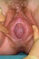
This shows the names of the parts of the vulva. The rest of the illustrations do not have labels.

This is a perfect annular hymen. It is called annular because the hymen forms a ring around the vaginal opening. As the hymen starts to erode from sexual or other activity, the hymen becomes less ring-like.

This is a crescentic, or lunar, hymen. It forms a crescent shape, like a half moon, above or (as in this case) below the vaginal opening.

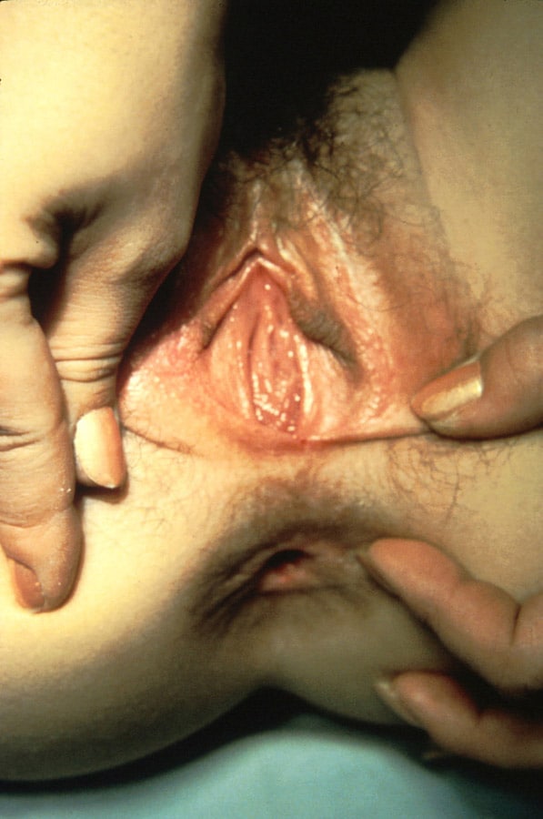
The hymen of a female with some sexual or masturbatory (internal) experience is apt to look something like this. Note that it is much less ring-like than the annular hymen.

This is what the hymen of a female who has only had a small amount of sexual activity or object insertion would look like. Health professionals who examine hymens for signs of sexual abuse are usually most interested in the posterior part of the hymen, from the 3 o'clock to 9 o'clock position. This is normally where the hymen breaks when the vagina is first penetrated.

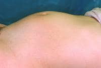
This is the vulva of a woman who has given birth. The hymen is completely gone, or nearly so.

One in 2000 girls is born with an imperforate hymen. A doctor will do surgery to create a hole in the hymen of such a newborn.

This is a rare cribriform hymen, characterized by many small holes. This type of hymen lets menstrual and other fluids out with no problem, but sexual activity and the insertion of tampons can be problematic.

This is a rare denticular hymen, so called because it looks like a set of teeth surrounding the vaginal opening.

This is a rare fimbriated hymen, with an irregular pattern around the vaginal opening.

This rare labial hymen looks like a third set of vulvar lips.

Some girls are born with only a tiny hole in their hymens. Surgery is also necessary for these newborns to create a larger vaginal opening.

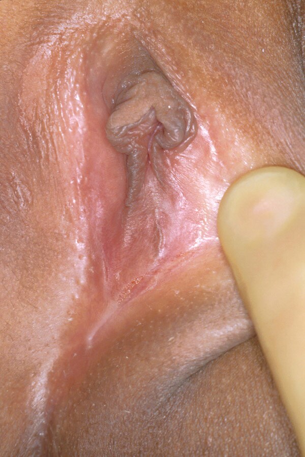
This rarity is called a septate hymen because of the piece of hymen that makes a septum, or bridge, across the vaginal opening.

This is the rare subseptate hymen, similar to the septate hymen only not making a bridge all the way across. Doesn't this remind you of the view into your throat with the uvula hanging down?


On painful first-time sex and the 'myths' of horror stories


First-time sex should not be painful
Hymens are not supposed to break…
and not all are supposed to bleed...


We do not believe that nature or God or the Goddess created us women with such faulty bodies that half of the things which should come natural to us, such as first-time sex or menstruation etc, are often painful instead.

We suspect that men's ignorance and intolerance unfortunately brought a lot of unnecessary and unnatural pain to us and one is the pain a lot of girls still go through the first time they have sex.

It is believed by many women and men that hymens will HAVE TO ‘break’ during first-time sex and that pain and/blood will be a common experience.
Now, we are NOT saying that it is a myth that for many young girls first-time sex will be a horror story and painful experience with blood, pain and discomfort. We know that those myths aren't myths at all. Unfortunately painful first-time sex does happen, those horror stories aren't fiction, they are vivid recollections of a traumatic experience.
And it happens way too often than it should.
So that is not the myth we are referring to in our title. What is a myth is that this HAS to happen.
What we are saying is that it is NOT natural at all for things to go that way...


THINGS SHOULD NOT GO THAT WAY.
The myth is the wrong belief that some young girls hold, that it will be up to chance or luck only whether or not their first time sex will be painful or not, and whether or not their hymen will break bloodlessly or not break at all.
It is not up to luck! Or it shouldn't be..!
That is the myth we are trying hard to break..
So the truth (compared to the myth) is that:

IF there was better information about first-time sex NOT having to hurt
IF there was more information for girls on how to stretch a hymen painlessly
IF boys were raised differently about sex and STOPPED thrusting as soon as things got discomfortable or painful for the girl they are having sex with for the first time
IF more women were taught about their hymen
If there was more information on vaginismus

then sex would NOT be a horror story or a painful experience the first time EVER.

Of course some women will experience a little pain, no matter what, and many will feel a twinge. It is, after all, a pretty big physical adjustment. But we should distinguish between feeling a little soreness or feelings of "stretching" and real pain. It should not be excruciating.
Beware of those who say you HAVE TO put up with some of it. There is NO reason you should put up with ANY pain!
Or is there? Why would you want to put up with pain or a man want you to put up with pain?
Ask yourself if you are under any pressure there and how you feel about it. If you were the man, would you want your partner to experience a lot of pain during your first sexual encounter ?
Plus, it could and does cause vaginismus to put up with pain during sex ( first time or not), so why risk it...?
We believe that no partner who is there with you should have you put up with any pain if he's a 'real man' (whatever that means..) and that you shouldn't just put up with him to show him you're a woman either. If there is pain, the boy/man should stop. Period. It means you are not ready yet evidently..

Plus, if there is pain, there may be other issues which again shouldn't be ignored. For instance:
the woman isn't lubricated enough her first time? Well, there's no reason that needs to happen. If she doesn't lubricate enough naturally there are artificial lubes which work just as well. If she isn't lubricated enough because she isn't aroused enough, well, then why is she having sex at all?


Sex is not a rite of passage or certainly shouldn't be.
Sex doesn't turn a girl into a woman or a boy into a man, how could it ?
Love probably does... Compassion, empathy, responsibility, consciousness, knowledge.. Not intercourse..

How to avoid painful first-time sex and the truth on 'breaking the hymen'

Now, one one way to avoid this is simply that of stretching the hymen yourself (or with the help of your partner) little by little, until it’s all gone.
It doesn't mean you have to use dilators, your hands or your partners' hands can be all that you need and it can be a beautiful, gentle, loving, erotic experience for both of you too.

Now, for some young girls, the idea of using a dilator or even her finger to stretch her hymen will sound unappealing.. But why? why not try using a dilator before you have sex for the first time? It's your vagina, why shouldn't you explore it in as many ways as possible before you share it with a partner? If it were ever suggested that a boy should keep a part of his body completely mysterious to himself and unexplored until his first sexual encounter with a woman, because it was a "rite of passage", it would be rightfully treated as backward nonsense.

Many girls would be uncomfortable about the idea of experimenting with any kind of penetration before intercourse, and many will be terribly afraid of no longer having a hymen because their whole reputation is based on whether or not they are virgins and marriagable in some countries, but that's because they've been raised to view their bodies as something they need to keep untouched for a man, which is what all these notions of "purity" and "virginity" are really about.
But a woman's body belongs to the woman!

She can and should be encouraged to explore it on her own terms. We are repulsed by the idea that first-time sex should be about "gritting your teeth" and bearing it, and until more people start seeing the flaws in this type of thinking and start teaching girls properly, it's going to keep on being about that.

As we have seen, there are a lot of misconceptions and much ignorance about the hymen and this can understandably become a cause of primary vaginismus because the muscles around the vagina will in fact clamp anticipating a painful first insertion. So if you heard horror stories about first-time sex, we hope reading this and the following links will reassure you and this may prevent vaginismus, so spread the good news! :)

Hopefully the next explanations and advice will help you clarify some doubts.
If not, but you may find the pictures too graphic there, so you are warned.

Ok, here we go:

There is wide range of experiences among women with their hymens so there could be exceptions to the following general descriptions.

In general, most hymens can be gently and gradually stretched with dilators or fingers until they (painlessly) widen. A lot of women find that their hymens pull apart so easily that they don't even notice it happening. No blood and no pain whatsoever…

If you gently stretch it yourself, a little bit at a time, (over days or weeks too, not necessarily in one session!) and if you do it with your smallest finger or your partner's smallest finger, you won’t even notice how it disappears. It's not dramatic at all and shouldn’t be. It's a gradual process.
If you're in control of the stretching, it can be done very gently.

Other hymens may be a bit thicker instead and when they pull apart, sometimes you may experience a tiny, sharp pain and you may see a tiny drop of blood coming out but very small numbers of women bleed more than that. If you stretch gently and constantly, bleeding is not at all common (but if it should happen, don’t worry, it happens to some).

The myth that hymens will break and that it will be very painful and bloody is certainly one of the causes of primary vaginismus for many young girls, so if we want to prevent vaginismus from happening to women in the future generations, or to avoid many future hymenectomies or painful first sexual experiences, we can help them by spreading the truth about hymens and teach our daughters, sisters, best friends etc. how they can stretch their own hymen painlessly over time.
It can be a beautiful empowering experience.

Boys too will need to hear that girls’ hymens are not made for them to break them… They are fine the way they are and already have 'holes'. They will just need to help their partners dilate gradually with a clean finger, (cut the nails!) to the point the woman is comfortable with inserting a penis, but for NO reason they should think that it WILL take a bit of ‘aggression’ and some tearing or that that's the NORMAL thing to happen or that the young woman will HAVE TO put up with some pain.

Again: we think there is NO reason you should put up with any pain at your first sexual experience!
Too many young lads grow up thinking that’s what’s supposed to happen… Only an inexperienced, rough boy coupled with a girl who does not know much about her hymen or who has a pretty thick one would make for a painful first time sexual experience.

With more knowledge and more compassion, thankfully in the future women won’t have to be afraid of their first time anymore or have terribly shocking first wedding nights...


Hymens, Hymenectomies and Vaginismus

For most women, hymens are perfectly fine the way they are and will not be the cause of vaginismus nor the obstacle to having painfree i/c or to dilate.
It is very rare for hymens to be totally closed (imperforated hymen) and that is usually detected very early on in life, cause it leads to an inability to menstruate. Estimates of an imperforated hymen frequency vary from 1 case per 1000 population to 1 case per 10,000 population.

So if you menstruate or have managed to insert a tampon or finger or anything in your vagina at least once, it is very unlikely that your hymen will need to be surgically opened, as you clearly have some kind of space there, so it may just need a bit of stretching instead, rather than an operation.

We found in our experience that gynecologists seem too eager to cut every hymen that's even a little bit problematic and they may not tell you that by no means is the surgery an instant cure for vaginismus. Also, the surgery isn't exactly comfortable, so you may associate pain with your genitals, which can complicate matters more.

There are a lot of misunderstandings about hymens and these can have caused you a lot of anxiety and lead you to require or accept a hymenectomy without careful considerations.

For instance, you may be terrified of tearing the hymen yourself, you may fear that it could be so painful that you would not be able to stand it. So with the hymen gone, you may think you’ll be able to insert dilators and a penis without that extra fear. But remember that even with the hymen out of the way, you will still have the PC muscle to contend with. Unlike the hymen, these muscles are located inside the vagina, about an inch or two around the vaginal entrance. They can still clamp in anticipation of pain so you will still need to gently retrain them in most cases, by gradual dilating,.

So, before deciding whether or not to go through with a hymenectomy, just make sure you don’t have the wrong expectations. We suggest you get to check the section on the Vulvar Anatomy too, that's very important to get comfortable with the whole area, so you will get to know your hymen well first and then see if it can really just need a little help from you.

Clues that you may really benefit from a hymenectomy to solve vaginismus


Remember: in MOST cases, a hymenectomy does not really seem necessary and we heard from many women with vag. who after that operation still had problems inserting dilators or having sex. As we explained, that's because the muscles could still clamp. However:

*If you have no fear or anxiety regarding sex and penetration and you feel as relaxed as can be during foreplay, yet you or your partner can’t get past the hymen, maybe your hymen may indeed be more to blame than your muscles, so a hymenectomy could be a good idea.

**If you are an athlete, you may have a hymen ‘band’ , basically you will have a regular small hole in your hymen, but the rest of the hymeneal skin around it may have become inelastic and quite rigid and you may find it impossible to insert a tampon.

**If you or your gynaecologist could not insert a q-tip, during her/his attempts at examination, or if that hurt a lot, and if they diagnosed you with “introitus-stenosis”, then you may benefit from this operation.

***But the best deciding factor is probably the level of pain you feel. If your hymen is really tender, thick and painful when you try to insert the smallest dilator or finger in it, or when you try stretching it, then it wouldn't be so good to put yourself through a lot of agony.

So you may first give yourself some time to think about it and in the meantime you could try and see if your hymen will slowly pull apart with some gentle stretching..

If you still believe that the surgery would be helpful to you, and if you are aware of the expectations you may have about it, then go for it by all means but remember that it’s not an immediate treatment for vaginismus.

Also, since any surgery with anaesthesia is not to be taken lightly, we advise you to first get a second opinion.